 Movie
Movie Controller


+ Open data

- Basic information
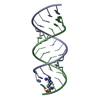
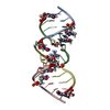
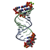
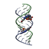
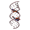
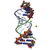
| Entry | Database: PDB / ID: 2jlt | ||||||
|---|---|---|---|---|---|---|---|
| Title | Crystal structure of an RNA kissing complex | ||||||
 Components Components |
| ||||||
 Keywords Keywords |  RNA / KISSING COMPLEX RNA / KISSING COMPLEX | ||||||
| Function / homology | RNA / RNA (> 10) Function and homology information Function and homology information | ||||||
| Biological species |  ESCHERICHIA COLI (E. coli) ESCHERICHIA COLI (E. coli) | ||||||
| Method | X-RAY DIFFRACTION / SYNCHROTRON / SAD / Resolution: 2.9 Å | ||||||
 Authors Authors | DiPrimo, C. / Fribourg, S. | ||||||
 Citation Citation | Journal: Nucleic Acids Res. / Year: 2008 Title: Exploring Tar-RNA Aptamer Loop-Loop Interaction by X-Ray Crystallography, Uv Spectroscopy and Surface Plasmon Resonance. Authors: Lebars, I. / Legrand, P. / Aime, A. / Pinaud, N. / Fribourg, S. / Di Primo, C. | ||||||
| History |
|
- Structure visualization
| Structure viewer | Molecule: MolmilJmol/JSmol |
|---|
- Downloads & links
-Download
| PDBx/mmCIF format | 2jlt.cif.gz | 28 KB | Display | PDBx/mmCIF format |
|---|---|---|---|---|
| PDB format | pdb2jlt.ent.gz | 19.3 KB | Display | PDB format |
| PDBx/mmJSON format | 2jlt.json.gz | Tree view | PDBx/mmJSON format | |
| Others |  Other downloads Other downloads |
-Validation report
| Arichive directory | https://data.pdbj.org/pub/pdb/validation_reports/jl/2jltftp://data.pdbj.org/pub/pdb/validation_reports/jl/2jlt | HTTPS FTP |
|---|
-Related structure data
| Similar structure data |
|---|


-Links
 PDBj
PDBj


- Assembly
| Deposited unit | 
| ||||||||
|---|---|---|---|---|---|---|---|---|---|
| 1 |
| ||||||||
| Unit cell |
|
-Components
| #1: RNA chain | Mass: 5841.395 Da / Num. of mol.: 1 / Source method: obtained synthetically / Source: (synth.) ESCHERICHIA COLI (E. coli) |
|---|---|
| #2: RNA chain | Mass: 5247.030 Da / Num. of mol.: 1 / Source method: obtained synthetically / Source: (synth.) ESCHERICHIA COLI (E. coli) |
-Experimental details
-Experiment
| Experiment | Method: X-RAY DIFFRACTION / Number of used crystals: 1 |
|---|
- Sample preparation
| Crystal | Density Matthews: 2.28 Å3/Da / Density % sol: 46 % / Description: NONE |
|---|---|
| Crystal grow | pH: 4.6 / Details: NA CITRATE 100MM, MPD 35%, pH 4.6 |
-Data collection
| Diffraction | Mean temperature: 100 K |
|---|---|
| Diffraction source | Source: SYNCHROTRON / Site: ESRF  / Beamline: BM14 / Wavelength: 0.91 / Beamline: BM14 / Wavelength: 0.91 |
| Detector | Type: MARRESEARCH / Detector: CCD / Date: Mar 3, 2007 |
| Radiation | Protocol: SINGLE WAVELENGTH / Monochromatic (M) / Laue (L): M / Scattering type: x-ray |
| Radiation wavelength | Wavelength: 0.91 Å / Relative weight: 1 |
| Reflection | Resolution: 2.9→20 Å / Num. obs: 10330 / % possible obs: 98.9 % / Observed criterion σ(I): 2 / Redundancy: 6.43 % / Rmerge(I) obs: 0.08 / Net I/σ(I): 18.66 |
| Reflection shell | Resolution: 2.9→3.07 Å / Redundancy: 6.09 % / Rmerge(I) obs: 0.38 / Mean I/σ(I) obs: 5.45 / % possible all: 96.6 |
- Processing
| Software |
| ||||||||||||||||||||||||||||||||||||||||||||||||||||||||||||||||||||||||||||||||||||||||||||||||||||||||||||||||||||||||||||||||||||||||||||||||||||||||||||||||||||||||||||||||||||||
|---|---|---|---|---|---|---|---|---|---|---|---|---|---|---|---|---|---|---|---|---|---|---|---|---|---|---|---|---|---|---|---|---|---|---|---|---|---|---|---|---|---|---|---|---|---|---|---|---|---|---|---|---|---|---|---|---|---|---|---|---|---|---|---|---|---|---|---|---|---|---|---|---|---|---|---|---|---|---|---|---|---|---|---|---|---|---|---|---|---|---|---|---|---|---|---|---|---|---|---|---|---|---|---|---|---|---|---|---|---|---|---|---|---|---|---|---|---|---|---|---|---|---|---|---|---|---|---|---|---|---|---|---|---|---|---|---|---|---|---|---|---|---|---|---|---|---|---|---|---|---|---|---|---|---|---|---|---|---|---|---|---|---|---|---|---|---|---|---|---|---|---|---|---|---|---|---|---|---|---|---|---|---|---|
| Refinement | Method to determine structure: SAD Starting model: NONE Resolution: 2.9→60.08 Å / Cor.coef. Fo:Fc: 0.952 / Cor.coef. Fo:Fc free: 0.921 / SU B: 25.93 / SU ML: 0.205 / TLS residual ADP flag: LIKELY RESIDUAL / Cross valid method: THROUGHOUT / ESU R: 0.37 / ESU R Free: 0.269 / Stereochemistry target values: MAXIMUM LIKELIHOOD Details: HYDROGENS HAVE BEEN ADDED IN THE RIDING POSITIONS. U VALUES ARE RESIDUAL ONLY
| ||||||||||||||||||||||||||||||||||||||||||||||||||||||||||||||||||||||||||||||||||||||||||||||||||||||||||||||||||||||||||||||||||||||||||||||||||||||||||||||||||||||||||||||||||||||
| Solvent computation | Ion probe radii: 0.8 Å / Shrinkage radii: 0.8 Å / VDW probe radii: 1.4 Å / Solvent model: MASK | ||||||||||||||||||||||||||||||||||||||||||||||||||||||||||||||||||||||||||||||||||||||||||||||||||||||||||||||||||||||||||||||||||||||||||||||||||||||||||||||||||||||||||||||||||||||
| Displacement parameters | Biso mean: 26.54 Å2 | ||||||||||||||||||||||||||||||||||||||||||||||||||||||||||||||||||||||||||||||||||||||||||||||||||||||||||||||||||||||||||||||||||||||||||||||||||||||||||||||||||||||||||||||||||||||
| Refinement step | Cycle: LAST / Resolution: 2.9→60.08 Å
| ||||||||||||||||||||||||||||||||||||||||||||||||||||||||||||||||||||||||||||||||||||||||||||||||||||||||||||||||||||||||||||||||||||||||||||||||||||||||||||||||||||||||||||||||||||||
| Refine LS restraints |
|