 Movie
Movie Controller

[English] 日本語
 Yorodumi
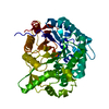
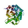
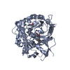
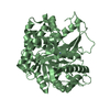
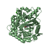
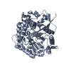
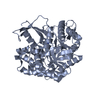
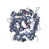
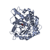
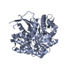
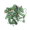
- PDB-2jie: BETA-GLUCOSIDASE B FROM BACILLUS POLYMYXA COMPLEXED WITH 2-F-GLUCOSE -
+ Open data

- Basic information
| Entry | Database: PDB / ID: 2jie | ||||||
|---|---|---|---|---|---|---|---|
| Title | BETA-GLUCOSIDASE B FROM BACILLUS POLYMYXA COMPLEXED WITH 2-F-GLUCOSE | ||||||
 Components Components | BETA-GLUCOSIDASE B | ||||||
 Keywords Keywords |  HYDROLASE / GLYCOSYL HYDROLASE FAMILY 1 / CARBOHYDRATE METABOLISM / POLYSACCHARIDE DEGRADATION / 2-F-GLUCOSE COMPLEX / CELLULOSE DEGRADATION / GLYCOSIDASE / BETA-GLUCOSIDASE HYDROLASE / GLYCOSYL HYDROLASE FAMILY 1 / CARBOHYDRATE METABOLISM / POLYSACCHARIDE DEGRADATION / 2-F-GLUCOSE COMPLEX / CELLULOSE DEGRADATION / GLYCOSIDASE / BETA-GLUCOSIDASE | ||||||
| Function / homology |  Function and homology information Function and homology informationscopolin beta-glucosidase activity / beta-glucosidase / beta-glucosidase activity / cellulose catabolic processSimilarity search - Function | ||||||
| Biological species |  PAENIBACILLUS POLYMYXA (bacteria) PAENIBACILLUS POLYMYXA (bacteria) | ||||||
| Method | X-RAY DIFFRACTION / SYNCHROTRON / MOLECULAR REPLACEMENT / Resolution: 2.3 Å | ||||||
 Authors Authors | Isorna, P. / Polaina, J. / Sanz-Aparicio, J. | ||||||
 Citation Citation | Journal: J.Mol.Biol. / Year: 2007 Title: Crystal Structures of Paenibacillus Polymyxa Beta-Glucosidase B Complexes Reveal the Molecular Basis of Substrate Specificity and Give New Insights Into the Catalytic Machinery of Family I Glycosidases. Authors: Isorna, P. / Polaina, J. / Latorre-Garcia, L. / Canada, F.J. / Gonzalez, B. / Sanz-Aparicio, J. | ||||||
| History |
| ||||||
| Remark 700 | SHEET DETERMINATION METHOD: DSSP THE SHEETS PRESENTED AS "AA" IN EACH CHAIN ON SHEET RECORDS BELOW ... SHEET DETERMINATION METHOD: DSSP THE SHEETS PRESENTED AS "AA" IN EACH CHAIN ON SHEET RECORDS BELOW IS ACTUALLY AN 9-STRANDED BARREL THIS IS REPRESENTED BY A 10-STRANDED SHEET IN WHICH THE FIRST AND LAST STRANDS ARE IDENTICAL. |
- Structure visualization
| Structure viewer | Molecule: MolmilJmol/JSmol |
|---|
- Downloads & links
-Download
| PDBx/mmCIF format | 2jie.cif.gz | 104.8 KB | Display | PDBx/mmCIF format |
|---|---|---|---|---|
| PDB format | pdb2jie.ent.gz | 79.5 KB | Display | PDB format |
| PDBx/mmJSON format | 2jie.json.gz | Tree view | PDBx/mmJSON format | |
| Others |  Other downloads Other downloads |
-Validation report
| Arichive directory | https://data.pdbj.org/pub/pdb/validation_reports/ji/2jieftp://data.pdbj.org/pub/pdb/validation_reports/ji/2jie | HTTPS FTP |
|---|
-Related structure data
| Related structure data |  2o9pC 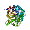 2o9rC  2o9tC  2z1sC  1e4iS S: Starting model for refinement C: citing same article ( |
|---|---|
| Similar structure data |


-Links
 PDBj
PDBj- Assembly
| Deposited unit | 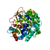
| ||||||||
|---|---|---|---|---|---|---|---|---|---|
| 1 |
| ||||||||
| Unit cell |
|
-Components
| #1: Protein | Mass: 52457.844 Da / Num. of mol.: 1 / Fragment: RESIDUES 2-448 Source method: isolated from a genetically manipulated source Source: (gene. exp.) PAENIBACILLUS POLYMYXA (bacteria) / Plasmid: DPUC18 / Production host: ESCHERICHIA COLI (E. coli) / Strain (production host): XL1BLUE / References: UniProt: P22505, beta-glucosidase |
|---|---|
| #2: Sugar | ChemComp-G2F /   Type: D-saccharide, alpha linking / Mass: 182.147 Da / Num. of mol.: 1 Type: D-saccharide, alpha linking / Mass: 182.147 Da / Num. of mol.: 1Source method: isolated from a genetically manipulated source Formula: C6H11FO5 |
| #3: Water | ChemComp-HOH / Water Mass: 18.015 Da / Num. of mol.: 129 / Source method: isolated from a natural source / Formula: H2O Mass: 18.015 Da / Num. of mol.: 129 / Source method: isolated from a natural source / Formula: H2O |
| Sequence details | HIS TAG |
-Experimental details
-Experiment
| Experiment | Method: X-RAY DIFFRACTION / Number of used crystals: 1 |
|---|
- Sample preparation
| Crystal | Density Matthews: 2.6 Å3/Da / Density % sol: 40 % / Description: NONE |
|---|
-Data collection
| Diffraction | Mean temperature: 120 K |
|---|---|
| Diffraction source | Source: SYNCHROTRON / Site: ESRF  / Beamline: BM16 / Wavelength: 0.9 / Beamline: BM16 / Wavelength: 0.9 |
| Detector | Type: MARRESEARCH / Detector: CCD |
| Radiation | Protocol: SINGLE WAVELENGTH / Monochromatic (M) / Laue (L): M / Scattering type: x-ray |
| Radiation wavelength | Wavelength: 0.9 Å / Relative weight: 1 |
| Reflection | Resolution: 2.2→25 Å / Num. obs: 24641 / % possible obs: 99.8 % / Observed criterion σ(I): 2 / Redundancy: 10.4 % / Biso Wilson estimate: 44.16 Å2 / Rmerge(I) obs: 0.09 / Net I/σ(I): 16 |
| Reflection shell | Resolution: 2.2→2.32 Å / Redundancy: 10.7 % / Rmerge(I) obs: 0.38 / Mean I/σ(I) obs: 3.4 / % possible all: 100 |
- Processing
| Software |
| ||||||||||||||||||||||||||||||||||||||||||||||||||||||||||||
|---|---|---|---|---|---|---|---|---|---|---|---|---|---|---|---|---|---|---|---|---|---|---|---|---|---|---|---|---|---|---|---|---|---|---|---|---|---|---|---|---|---|---|---|---|---|---|---|---|---|---|---|---|---|---|---|---|---|---|---|---|---|
| Refinement | Method to determine structure: MOLECULAR REPLACEMENT Starting model: PDB ENTRY 1E4I Resolution: 2.3→25 Å / Data cutoff high absF: 10000 / Isotropic thermal model: RESTRAINED / Cross valid method: THROUGHOUT / σ(F): 2
| ||||||||||||||||||||||||||||||||||||||||||||||||||||||||||||
| Solvent computation | Solvent model: RESTRAINED / Bsol: 51.5349 Å2 / ksol: 0.38643 e/Å3 | ||||||||||||||||||||||||||||||||||||||||||||||||||||||||||||
| Displacement parameters |
| ||||||||||||||||||||||||||||||||||||||||||||||||||||||||||||
| Refinement step | Cycle: LAST / Resolution: 2.3→25 Å
| ||||||||||||||||||||||||||||||||||||||||||||||||||||||||||||
| Refine LS restraints |
| ||||||||||||||||||||||||||||||||||||||||||||||||||||||||||||
| LS refinement shell | Resolution: 2.3→2.33 Å / Total num. of bins used: 29 /
| ||||||||||||||||||||||||||||||||||||||||||||||||||||||||||||
| Xplor file |
|
