 Movie
Movie Controller

[English] 日本語
 Yorodumi
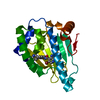
- PDB-2jfs: Crystal structure of the PPM Ser-Thr phosphatase MsPP from Mycoba... -
+ Open data

- Basic information
| Entry | Database: PDB / ID: 2jfs | ||||||
|---|---|---|---|---|---|---|---|
| Title | Crystal structure of the PPM Ser-Thr phosphatase MsPP from Mycobacterium smegmatis in complex with cacodylate | ||||||
 Components Components | SER-THR PHOSPHATASE MSPP | ||||||
 Keywords Keywords |  HYDROLASE / PPM PHOSPHATASE / MANGANESE / CACODYLATE / MYCOBACTERIUM HYDROLASE / PPM PHOSPHATASE / MANGANESE / CACODYLATE / MYCOBACTERIUM | ||||||
| Function / homology |  Function and homology informationprotein serine/threonine phosphatase activity / protein-serine/threonine phosphatase / metal ion binding Function and homology informationprotein serine/threonine phosphatase activity / protein-serine/threonine phosphatase / metal ion bindingSimilarity search - Function | ||||||
| Biological species |  MYCOBACTERIUM SMEGMATIS (bacteria) MYCOBACTERIUM SMEGMATIS (bacteria) | ||||||
| Method | X-RAY DIFFRACTION / SYNCHROTRON / MOLECULAR REPLACEMENT / Resolution: 1.45 Å | ||||||
 Authors Authors | Bellinzoni, M. / Wehenkel, A. / Shepard, W. / Alzari, P.M. | ||||||
 Citation Citation | Journal: Structure / Year: 2007 Title: Insights Into the Mechanism of Ppm Ser/Thr Phosphatases from the Atomic Resolution Structures of a Mycobacterial Enzyme Authors: Bellinzoni, M. / Wehenkel, A. / Shepard, W. / Alzari, P.M. | ||||||
| History |
| ||||||
| Remark 700 | SHEET THE SHEET STRUCTURE OF THIS MOLECULE IS BIFURCATED. IN ORDER TO REPRESENT THIS FEATURE IN ... SHEET THE SHEET STRUCTURE OF THIS MOLECULE IS BIFURCATED. IN ORDER TO REPRESENT THIS FEATURE IN THE SHEET RECORDS BELOW, TWO SHEETS ARE DEFINED. |
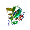
- Structure visualization
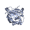
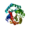
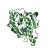
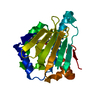
| Structure viewer | Molecule: MolmilJmol/JSmol |
|---|
- Downloads & links
-Download
| PDBx/mmCIF format | 2jfs.cif.gz | 66.2 KB | Display | PDBx/mmCIF format |
|---|---|---|---|---|
| PDB format | pdb2jfs.ent.gz | 47.6 KB | Display | PDB format |
| PDBx/mmJSON format | 2jfs.json.gz | Tree view | PDBx/mmJSON format | |
| Others |  Other downloads Other downloads |
-Validation report
| Arichive directory | https://data.pdbj.org/pub/pdb/validation_reports/jf/2jfsftp://data.pdbj.org/pub/pdb/validation_reports/jf/2jfs | HTTPS FTP |
|---|
-Related structure data
| Related structure data |  2jfrC  2jftC  2cm1S C: citing same article ( S: Starting model for refinement |
|---|---|
| Similar structure data |


-Links
 PDBj
PDBj


- Assembly
| Deposited unit | 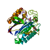
| ||||||||
|---|---|---|---|---|---|---|---|---|---|
| 1 |
| ||||||||
| Unit cell |
|
-Components
| #1: Protein | Mass: 24278.018 Da / Num. of mol.: 1 Source method: isolated from a genetically manipulated source Source: (gene. exp.) MYCOBACTERIUM SMEGMATIS (bacteria) / Strain: MC2 155 / Production host: ESCHERICHIA COLI (E. coli) / Strain (production host): BL21(DE3) / References: UniProt: A0QTQ6 | ||||||||
|---|---|---|---|---|---|---|---|---|---|
| #2: Chemical |   Mass: 54.938 Da / Num. of mol.: 3 / Source method: obtained synthetically / Formula: Mn Mass: 54.938 Da / Num. of mol.: 3 / Source method: obtained synthetically / Formula: Mn#3: Chemical | ChemComp-CAC / | Cacodylic acid  Mass: 136.989 Da / Num. of mol.: 1 / Source method: obtained synthetically / Formula: C2H6AsO2 Mass: 136.989 Da / Num. of mol.: 1 / Source method: obtained synthetically / Formula: C2H6AsO2#4: Chemical | Glycerol  Mass: 92.094 Da / Num. of mol.: 3 / Source method: obtained synthetically / Formula: C3H8O3 Mass: 92.094 Da / Num. of mol.: 3 / Source method: obtained synthetically / Formula: C3H8O3#5: Water | ChemComp-HOH / | Water Mass: 18.015 Da / Num. of mol.: 240 / Source method: isolated from a natural source / Formula: H2O Mass: 18.015 Da / Num. of mol.: 240 / Source method: isolated from a natural source / Formula: H2OSequence details | FIRST RESIDUE GLY IS A CLONING ARTIFACT | |
-Experimental details
-Experiment
| Experiment | Method: X-RAY DIFFRACTION / Number of used crystals: 1 |
|---|
- Sample preparation
| Crystal | Density Matthews: 2.7 Å3/Da / Density % sol: 54.6 % |
|---|---|
| Crystal grow | pH: 6.5 Details: 18% PEG12K, 85 MM NA CACODYLATE PH 6.5, 120 MM NA ACETATE, 13% GLYCEROL |
-Data collection
| Diffraction | Mean temperature: 100 K |
|---|---|
| Diffraction source | Source: SYNCHROTRON / Site: ESRF  / Beamline: ID14-2 / Wavelength: 0.933 / Beamline: ID14-2 / Wavelength: 0.933 |
| Detector | Type: ADSC CCD / Detector: CCD / Date: Nov 7, 2005 / Details: MIRRORS |
| Radiation | Monochromator: GE(220) / Protocol: SINGLE WAVELENGTH / Monochromatic (M) / Laue (L): M / Scattering type: x-ray |
| Radiation wavelength | Wavelength: 0.933 Å / Relative weight: 1 |
| Reflection | Resolution: 1.45→25.9 Å / Num. obs: 47486 / % possible obs: 99 % / Redundancy: 3.8 % / Rmerge(I) obs: 0.05 / Net I/σ(I): 15.6 |
| Reflection shell | Resolution: 1.45→1.53 Å / Redundancy: 3.7 % / Rmerge(I) obs: 0.43 / Mean I/σ(I) obs: 2.6 / % possible all: 97.7 |
- Processing
| Software |
| ||||||||||||||||||||||||||||||||||||||||||||||||||||||||||||||||||||||||||||||||||||||||||||||||||||||||||||||||||||||||||||||||||||||||||||||||||||||||||||||||||||||||||||||||||||||
|---|---|---|---|---|---|---|---|---|---|---|---|---|---|---|---|---|---|---|---|---|---|---|---|---|---|---|---|---|---|---|---|---|---|---|---|---|---|---|---|---|---|---|---|---|---|---|---|---|---|---|---|---|---|---|---|---|---|---|---|---|---|---|---|---|---|---|---|---|---|---|---|---|---|---|---|---|---|---|---|---|---|---|---|---|---|---|---|---|---|---|---|---|---|---|---|---|---|---|---|---|---|---|---|---|---|---|---|---|---|---|---|---|---|---|---|---|---|---|---|---|---|---|---|---|---|---|---|---|---|---|---|---|---|---|---|---|---|---|---|---|---|---|---|---|---|---|---|---|---|---|---|---|---|---|---|---|---|---|---|---|---|---|---|---|---|---|---|---|---|---|---|---|---|---|---|---|---|---|---|---|---|---|---|
| Refinement | Method to determine structure: MOLECULAR REPLACEMENT Starting model: PDB ENTRY 2CM1 Resolution: 1.45→25.38 Å / Cor.coef. Fo:Fc: 0.968 / Cor.coef. Fo:Fc free: 0.965 / SU B: 1.93 / SU ML: 0.043 / TLS residual ADP flag: LIKELY RESIDUAL / Cross valid method: THROUGHOUT / ESU R: 0.062 / ESU R Free: 0.06 / Stereochemistry target values: MAXIMUM LIKELIHOOD Details: HYDROGENS HAVE BEEN ADDED IN THE RIDING POSITIONS. ATOM RECORD CONTAINS RESIDUAL B FACTORS ONLY
| ||||||||||||||||||||||||||||||||||||||||||||||||||||||||||||||||||||||||||||||||||||||||||||||||||||||||||||||||||||||||||||||||||||||||||||||||||||||||||||||||||||||||||||||||||||||
| Solvent computation | Ion probe radii: 0.8 Å / Shrinkage radii: 0.8 Å / VDW probe radii: 1.4 Å / Solvent model: MASK | ||||||||||||||||||||||||||||||||||||||||||||||||||||||||||||||||||||||||||||||||||||||||||||||||||||||||||||||||||||||||||||||||||||||||||||||||||||||||||||||||||||||||||||||||||||||
| Displacement parameters | Biso mean: 19.59 Å2
| ||||||||||||||||||||||||||||||||||||||||||||||||||||||||||||||||||||||||||||||||||||||||||||||||||||||||||||||||||||||||||||||||||||||||||||||||||||||||||||||||||||||||||||||||||||||
| Refinement step | Cycle: LAST / Resolution: 1.45→25.38 Å
| ||||||||||||||||||||||||||||||||||||||||||||||||||||||||||||||||||||||||||||||||||||||||||||||||||||||||||||||||||||||||||||||||||||||||||||||||||||||||||||||||||||||||||||||||||||||
| Refine LS restraints |
|