 Movie
Movie Controller

[English] 日本語
 Yorodumi
- PDB-2j5k: 2.0 A resolution structure of the wild type malate dehydrogenase ... -
+ Open data

- Basic information
| Entry | Database: PDB / ID: 2j5k | ||||||
|---|---|---|---|---|---|---|---|
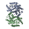
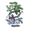
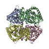
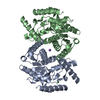
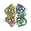
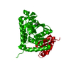
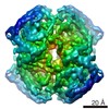
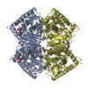
| Title | 2.0 A resolution structure of the wild type malate dehydrogenase from Haloarcula marismortui (radiation damage series) | ||||||
 Components Components | MALATE DEHYDROGENASE | ||||||
 Keywords Keywords | OXIDOREDUCTASE / NAD / HALOPHILE / RADIATION DAMAGE / MALATE DEHYDROGENASE / TRICARBOXYLIC ACID CYCLE | ||||||
| Function / homology |  Function and homology informationmalate dehydrogenase / L-malate dehydrogenase activity / carboxylic acid metabolic process / tricarboxylic acid cycle / cytoplasm Function and homology informationmalate dehydrogenase / L-malate dehydrogenase activity / carboxylic acid metabolic process / tricarboxylic acid cycle / cytoplasmSimilarity search - Function | ||||||
| Biological species |  HALOARCULA MARISMORTUI (Halophile) HALOARCULA MARISMORTUI (Halophile) | ||||||
| Method | X-RAY DIFFRACTION / SYNCHROTRON / MOLECULAR REPLACEMENT / Resolution: 2 Å | ||||||
 Authors Authors | Fioravanti, E. / Vellieux, F.M.D. / Amara, P. / Madern, D. / Weik, M. | ||||||
 Citation Citation | Journal: J.Synchrotron Radiat. / Year: 2007 Title: Specific Radiation Damage to Acidic Residues and its Relation to Their Chemical and Structural Environment. Authors: Fioravanti, E. / Vellieux, F.M.D. / Amara, P. / Madern, D. / Weik, M. #1: Journal: J.Mol.Biol. / Year: 2003Title: The Oligomeric States of Haloarcula Marismortui Malate Dehydrogenase are Modulated by Solvent Components as Shown by Crystallographic and Biochemical Studies Authors: Irimia, A. / Ebel, C. / Madern, D. / Richard, S.B. / Cosenza, L.W. / Zaccai, G. / D Vellieux, F.M. #2: Journal: Biochemistry / Year: 2000Title: Halophilic Adaptation: Novel Solvent Protein Interactions Observed in the 2.9 And 2.6 A Resolution Structures of the Wild Type and a Mutant of Malate Dehydrogenase from Haloarcula Marismortui Authors: Richard, S.B. / Madern, D. / Garcin, E. / Zaccai, G. #3: Journal: Science / Year: 1995Title: Structural Features that Stabilize Halophilic Malate-Dehydrogenase from an Archaebacterium. Authors: Dym, O. / Mevarech, M. / Sussman, J.L. | ||||||
| History |
|
- Structure visualization
| Structure viewer | Molecule: MolmilJmol/JSmol |
|---|
- Downloads & links
-Download
| PDBx/mmCIF format | 2j5k.cif.gz | 252.8 KB | Display | PDBx/mmCIF format |
|---|---|---|---|---|
| PDB format | pdb2j5k.ent.gz | 203.8 KB | Display | PDB format |
| PDBx/mmJSON format | 2j5k.json.gz | Tree view | PDBx/mmJSON format | |
| Others |  Other downloads Other downloads |
-Validation report
| Arichive directory | https://data.pdbj.org/pub/pdb/validation_reports/j5/2j5kftp://data.pdbj.org/pub/pdb/validation_reports/j5/2j5k | HTTPS FTP |
|---|
-Related structure data
| Related structure data |  2j5qC  2j5rC 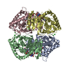 1o6zS S: Starting model for refinement C: citing same article ( |
|---|---|
| Similar structure data |


-Links
 PDBj
PDBj


- Assembly
| Deposited unit | 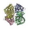
| ||||||||
|---|---|---|---|---|---|---|---|---|---|
| 1 |
| ||||||||
| Unit cell |
|
-Components
| #1: Protein | Mass: 32837.727 Da / Num. of mol.: 4 Source method: isolated from a genetically manipulated source Source: (gene. exp.) HALOARCULA MARISMORTUI (Halophile) / Strain: HMS174 / Plasmid: PET11A / Production host:  ESCHERICHIA COLI (E. coli) / Strain (production host): BL21(DE3) / References: UniProt: Q07841, malate dehydrogenase ESCHERICHIA COLI (E. coli) / Strain (production host): BL21(DE3) / References: UniProt: Q07841, malate dehydrogenase#2: Chemical | ChemComp-CL / Chloride  Mass: 35.453 Da / Num. of mol.: 8 / Source method: obtained synthetically / Formula: Cl Mass: 35.453 Da / Num. of mol.: 8 / Source method: obtained synthetically / Formula: Cl#3: Water | ChemComp-HOH / | Water Mass: 18.015 Da / Num. of mol.: 1023 / Source method: isolated from a natural source / Formula: H2O Mass: 18.015 Da / Num. of mol.: 1023 / Source method: isolated from a natural source / Formula: H2O |
|---|
-Experimental details
-Experiment
| Experiment | Method: X-RAY DIFFRACTION / Number of used crystals: 1 |
|---|
- Sample preparation
| Crystal | Density Matthews: 3.19 Å3/Da / Density % sol: 61.11 % |
|---|---|
| Crystal grow | Method: vapor diffusion, sitting drop / pH: 7 Details: 3UL OF PROTEIN PLUS 4UL OF MPD WERE EQUILIBRATED AGAINST 58% MPD VIA THE SITTING DROP REVERSE VAPOUR DIFFUSION TECHNIQUE, pH 7.00 |
-Data collection
| Diffraction | Mean temperature: 100 K |
|---|---|
| Diffraction source | Source: SYNCHROTRON / Site: ESRF  / Beamline: ID23-1 / Wavelength: 0.939 / Beamline: ID23-1 / Wavelength: 0.939 |
| Detector | Type: ADSC CCD / Detector: CCD / Date: Mar 4, 2005 / Details: MIRRORS |
| Radiation | Monochromator: SILICON (1 1 1) CHANNEL- CUT / Protocol: SINGLE WAVELENGTH / Monochromatic (M) / Laue (L): M / Scattering type: x-ray |
| Radiation wavelength | Wavelength: 0.939 Å / Relative weight: 1 |
| Reflection | Resolution: 2→20 Å / Num. obs: 112033 / % possible obs: 93.9 % / Observed criterion σ(I): 0 / Redundancy: 3.2 % / Biso Wilson estimate: 12.851 Å2 / Rmerge(I) obs: 0.1 / Net I/σ(I): 11.1 |
| Reflection shell | Resolution: 2→2.11 Å / Redundancy: 3.2 % / Rmerge(I) obs: 0.3 / Mean I/σ(I) obs: 3 / % possible all: 85.4 |
- Processing
| Software |
| ||||||||||||||||||||||||||||||||||||||||||||||||||||||||||||||||||||||||||||||||
|---|---|---|---|---|---|---|---|---|---|---|---|---|---|---|---|---|---|---|---|---|---|---|---|---|---|---|---|---|---|---|---|---|---|---|---|---|---|---|---|---|---|---|---|---|---|---|---|---|---|---|---|---|---|---|---|---|---|---|---|---|---|---|---|---|---|---|---|---|---|---|---|---|---|---|---|---|---|---|---|---|---|
| Refinement | Method to determine structure: MOLECULAR REPLACEMENT Starting model: PDB ENTRY 1O6Z Resolution: 2→20 Å / Data cutoff high absF: 10000 / Isotropic thermal model: RESTRAINED / Cross valid method: THROUGHOUT / σ(F): 0 / Stereochemistry target values: MAXIMUM LIKELIHOOD Details: SIDE CHAINS ATOMS WITHOUT ELECTRON DENSITY APPEARING AT 1 SIGMA LEVEL IN 3MFO-2DFC MAPS WERE REMOVED FROM THE MODEL.RESIDUES IN POSITION 100-107 WERE EXCLUDED FROM THE MODEL OF MONOMERS B ...Details: SIDE CHAINS ATOMS WITHOUT ELECTRON DENSITY APPEARING AT 1 SIGMA LEVEL IN 3MFO-2DFC MAPS WERE REMOVED FROM THE MODEL.RESIDUES IN POSITION 100-107 WERE EXCLUDED FROM THE MODEL OF MONOMERS B AND D BECAUSE DISORDERED.
| ||||||||||||||||||||||||||||||||||||||||||||||||||||||||||||||||||||||||||||||||
| Solvent computation | Solvent model: FLAT MODEL / Bsol: 64.2269 Å2 / ksol: 0.380188 e/Å3 | ||||||||||||||||||||||||||||||||||||||||||||||||||||||||||||||||||||||||||||||||
| Displacement parameters | Biso mean: 21.3 Å2
| ||||||||||||||||||||||||||||||||||||||||||||||||||||||||||||||||||||||||||||||||
| Refine analyze |
| ||||||||||||||||||||||||||||||||||||||||||||||||||||||||||||||||||||||||||||||||
| Refinement step | Cycle: LAST / Resolution: 2→20 Å
| ||||||||||||||||||||||||||||||||||||||||||||||||||||||||||||||||||||||||||||||||
| Refine LS restraints |
| ||||||||||||||||||||||||||||||||||||||||||||||||||||||||||||||||||||||||||||||||
| LS refinement shell | Resolution: 2→2.07 Å / Total num. of bins used: 10
| ||||||||||||||||||||||||||||||||||||||||||||||||||||||||||||||||||||||||||||||||
| Xplor file |
|
