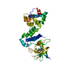
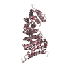
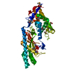
Entry Database : PDB / ID : 2j41Title Crystal structure of Staphylococcus aureus guanylate monophosphate kinase GUANYLATE KINASE Keywords / / / / / / Function / homology Biological species STAPHYLOCOCCUS AUREUS (bacteria)Method / / / Resolution : 1.9 Å Authors El Omari, K. / Dhaliwal, B. / Lockyer, M. / Charles, I. / Hawkins, A.R. / Stammers, D.K. Journal : Acta Crystallogr.,Sect.F / Year : 2006Title : Structure of Staphylococcus Aureus Guanylate Monophosphate KinaseAuthors : El Omari, K. / Dhaliwal, B. / Lockyer, M. / Charles, I. / Hawkins, A.R. / Stammers, D.K. History Deposition Aug 24, 2006 Deposition site / Processing site Revision 1.0 Oct 11, 2006 Provider / Type Revision 1.1 May 8, 2011 Group Revision 1.2 Jul 13, 2011 Group Revision 1.3 Dec 13, 2023 Group Data collection / Database references ... Data collection / Database references / Derived calculations / Other / Refinement description Category chem_comp_atom / chem_comp_bond ... chem_comp_atom / chem_comp_bond / database_2 / pdbx_database_status / pdbx_initial_refinement_model / pdbx_struct_conn_angle / struct_conn Item _database_2.pdbx_DOI / _database_2.pdbx_database_accession ... _database_2.pdbx_DOI / _database_2.pdbx_database_accession / _pdbx_database_status.status_code_sf / _pdbx_struct_conn_angle.ptnr1_auth_comp_id / _pdbx_struct_conn_angle.ptnr1_auth_seq_id / _pdbx_struct_conn_angle.ptnr1_label_asym_id / _pdbx_struct_conn_angle.ptnr1_label_atom_id / _pdbx_struct_conn_angle.ptnr1_label_comp_id / _pdbx_struct_conn_angle.ptnr1_label_seq_id / _pdbx_struct_conn_angle.ptnr3_auth_comp_id / _pdbx_struct_conn_angle.ptnr3_auth_seq_id / _pdbx_struct_conn_angle.ptnr3_label_asym_id / _pdbx_struct_conn_angle.ptnr3_label_atom_id / _pdbx_struct_conn_angle.ptnr3_label_comp_id / _pdbx_struct_conn_angle.ptnr3_label_seq_id / _pdbx_struct_conn_angle.value / _struct_conn.pdbx_dist_value / _struct_conn.ptnr1_auth_comp_id / _struct_conn.ptnr1_auth_seq_id / _struct_conn.ptnr1_label_asym_id / _struct_conn.ptnr1_label_atom_id / _struct_conn.ptnr1_label_comp_id / _struct_conn.ptnr1_label_seq_id / _struct_conn.ptnr2_auth_comp_id / _struct_conn.ptnr2_auth_seq_id / _struct_conn.ptnr2_label_asym_id / _struct_conn.ptnr2_label_atom_id / _struct_conn.ptnr2_label_comp_id / _struct_conn.ptnr2_label_seq_id
Show all Show less
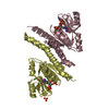
 Movie
Movie Controller

 Yorodumi
 Open data
 Basic information
 Components
 Keywords
 Function and homology information

 Authors
 Citation
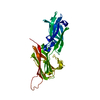
 Structure visualization
 Downloads & links
 Other downloads


 PDBj
PDBj

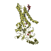
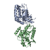
 Assembly
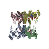


 Mass: 96.063 Da / Num. of mol.: 5 / Source method: obtained synthetically / Formula: SO4
 Mass: 39.098 Da / Num. of mol.: 3 / Source method: obtained synthetically / Formula: K
 Mass: 363.221 Da / Num. of mol.: 3 / Source method: obtained synthetically / Formula: C10H14N5O8P
 Mass: 18.015 Da / Num. of mol.: 471 / Source method: isolated from a natural source / Formula: H2O
 Sample preparation
 / Beamline: ID14-1 / Wavelength: 0.93
  Processing