 Movie
Movie Controller


+ Open data

- Basic information
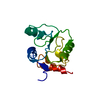
| Entry | Database: PDB / ID: 2j17 | ||||||
|---|---|---|---|---|---|---|---|
| Title | pTyr bound form of SDP-1 | ||||||
 Components Components | TYROSINE-PROTEIN PHOSPHATASE YIL113W | ||||||
 Keywords Keywords |  HYDROLASE / PROTEIN PHOSPHATASE / HYPOTHETICAL PROTEIN HYDROLASE / PROTEIN PHOSPHATASE / HYPOTHETICAL PROTEIN | ||||||
| Function / homology |  Function and homology information Function and homology informationcell wall integrity MAPK cascade / RAF-independent MAPK1/3 activation / ERKs are inactivated / Negative regulation of MAPK pathway / MAP kinase tyrosine phosphatase activity / protein tyrosine/threonine phosphatase activity / MAP kinase tyrosine/serine/threonine phosphatase activity / negative regulation of MAPK cascade / dephosphorylation / protein-tyrosine-phosphatase ...cell wall integrity MAPK cascade / RAF-independent MAPK1/3 activation / ERKs are inactivated / Negative regulation of MAPK pathway / MAP kinase tyrosine phosphatase activity / protein tyrosine/threonine phosphatase activity / MAP kinase tyrosine/serine/threonine phosphatase activity / negative regulation of MAPK cascade / dephosphorylation / protein-tyrosine-phosphatase / nucleus / cytoplasmSimilarity search - Function | ||||||
| Biological species |  SACCHAROMYCES CEREVISIAE (brewer's yeast) SACCHAROMYCES CEREVISIAE (brewer's yeast) | ||||||
| Method | X-RAY DIFFRACTION / SYNCHROTRON / MOLECULAR REPLACEMENT / Resolution: 2.84 Å | ||||||
 Authors Authors | Briggs, D.C. / McDonald, N.Q. | ||||||
 Citation Citation | Journal: Nature / Year: 2007 Title: Redox-mediated substrate recognition by Sdp1 defines a new group of tyrosine phosphatases. Authors: Fox, G.C. / Shafiq, M. / Briggs, D.C. / Knowles, P.P. / Collister, M. / Didmon, M.J. / Makrantoni, V. / Dickinson, R.J. / Hanrahan, S. / Totty, N. / Stark, M.J. / Keyse, S.M. / McDonald, N.Q. | ||||||
| History |
|
- Structure visualization
| Structure viewer | Molecule: MolmilJmol/JSmol |
|---|
- Downloads & links
-Download
| PDBx/mmCIF format | 2j17.cif.gz | 73.3 KB | Display | PDBx/mmCIF format |
|---|---|---|---|---|
| PDB format | pdb2j17.ent.gz | 53.4 KB | Display | PDB format |
| PDBx/mmJSON format | 2j17.json.gz | Tree view | PDBx/mmJSON format | |
| Others |  Other downloads Other downloads |
-Validation report
| Arichive directory | https://data.pdbj.org/pub/pdb/validation_reports/j1/2j17ftp://data.pdbj.org/pub/pdb/validation_reports/j1/2j17 | HTTPS FTP |
|---|
-Related structure data
| Related structure data |  2j16SC S: Starting model for refinement C: citing same article ( |
|---|---|
| Similar structure data |


-Links
 PDBj
PDBj


- Assembly
| Deposited unit | 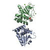
| |||||||||||||||||||||||||||||||||||||||||||||||||||||||||||
|---|---|---|---|---|---|---|---|---|---|---|---|---|---|---|---|---|---|---|---|---|---|---|---|---|---|---|---|---|---|---|---|---|---|---|---|---|---|---|---|---|---|---|---|---|---|---|---|---|---|---|---|---|---|---|---|---|---|---|---|---|
| 1 | 
| |||||||||||||||||||||||||||||||||||||||||||||||||||||||||||
| 2 | 
| |||||||||||||||||||||||||||||||||||||||||||||||||||||||||||
| Unit cell |
| |||||||||||||||||||||||||||||||||||||||||||||||||||||||||||
| Noncrystallographic symmetry (NCS) | NCS domain:
NCS domain segments: Ens-ID: 1 / Refine code: 6
NCS oper: (Code: given Matrix: (0.9605, -0.2211, -0.1688), Vector : |
-Components
| #1: Protein | Mass: 20853.939 Da / Num. of mol.: 2 / Fragment: RESIDUES 17-198 / Mutation: YES Source method: isolated from a genetically manipulated source Source: (gene. exp.) SACCHAROMYCES CEREVISIAE (brewer's yeast)Strain: S288C / AB972 / Plasmid: PET14B / Production host:  ESCHERICHIA COLI (E. coli) / Strain (production host): FB810 / Variant (production host): PLYSS / References: UniProt: P40479, protein-tyrosine-phosphatase ESCHERICHIA COLI (E. coli) / Strain (production host): FB810 / Variant (production host): PLYSS / References: UniProt: P40479, protein-tyrosine-phosphatase#2: Chemical | ChemComp-MG /   Mass: 24.305 Da / Num. of mol.: 5 / Source method: obtained synthetically / Formula: Mg Mass: 24.305 Da / Num. of mol.: 5 / Source method: obtained synthetically / Formula: Mg#3: Chemical |   Type: L-peptide linking / Mass: 261.168 Da / Num. of mol.: 2 / Source method: obtained synthetically / Formula: C9H12NO6P Type: L-peptide linking / Mass: 261.168 Da / Num. of mol.: 2 / Source method: obtained synthetically / Formula: C9H12NO6P#4: Water | ChemComp-HOH / | Water Mass: 18.015 Da / Num. of mol.: 24 / Source method: isolated from a natural source / Formula: H2O Mass: 18.015 Da / Num. of mol.: 24 / Source method: isolated from a natural source / Formula: H2OCompound details | ENGINEERED | |
|---|
-Experimental details
-Experiment
| Experiment | Method: X-RAY DIFFRACTION / Number of used crystals: 1 |
|---|
- Sample preparation
| Crystal | Density Matthews: 3.14 Å3/Da / Density % sol: 60.47 % |
|---|---|
| Crystal grow | Method: vapor diffusion, sitting drop Details: 28% PEG 400, 0.1M HEPES, PH 7.5, 0.2M CACL2, 7MG/ML PROTEIN, 15MM PHOSPHOTYROSINE, 1:1 SITTING DROP VAPOUR DIFFUSION |
-Data collection
| Diffraction | Mean temperature: 100 K |
|---|---|
| Diffraction source | Source: SYNCHROTRON / Site: SRS  / Beamline: PX14.2 / Wavelength: 0.97 / Beamline: PX14.2 / Wavelength: 0.97 |
| Detector | Type: ADSC CCD / Detector: CCD / Date: May 20, 2002 / Details: MIRRORS |
| Radiation | Monochromator: SI CRYSTAL / Protocol: SINGLE WAVELENGTH / Monochromatic (M) / Laue (L): M / Scattering type: x-ray |
| Radiation wavelength | Wavelength: 0.97 Å / Relative weight: 1 |
| Reflection | Resolution: 2.84→61.66 Å / Num. obs: 10632 / % possible obs: 94.7 % / Observed criterion σ(I): 1 / Redundancy: 3.8 % / Rmerge(I) obs: 0.08 / Net I/σ(I): 7.4 |
| Reflection shell | Resolution: 2.84→2.99 Å / Redundancy: 3.4 % / Rmerge(I) obs: 0.38 / Mean I/σ(I) obs: 1.9 / % possible all: 86.5 |
- Processing
| Software |
| ||||||||||||||||||||||||||||||||||||||||||||||||||||||||||||||||||||||||||||||||||||||||||||||||||||||||||||||||||||||||||||||||||||||||||||||||||||||||||||||||||||||||||||||||||||||
|---|---|---|---|---|---|---|---|---|---|---|---|---|---|---|---|---|---|---|---|---|---|---|---|---|---|---|---|---|---|---|---|---|---|---|---|---|---|---|---|---|---|---|---|---|---|---|---|---|---|---|---|---|---|---|---|---|---|---|---|---|---|---|---|---|---|---|---|---|---|---|---|---|---|---|---|---|---|---|---|---|---|---|---|---|---|---|---|---|---|---|---|---|---|---|---|---|---|---|---|---|---|---|---|---|---|---|---|---|---|---|---|---|---|---|---|---|---|---|---|---|---|---|---|---|---|---|---|---|---|---|---|---|---|---|---|---|---|---|---|---|---|---|---|---|---|---|---|---|---|---|---|---|---|---|---|---|---|---|---|---|---|---|---|---|---|---|---|---|---|---|---|---|---|---|---|---|---|---|---|---|---|---|---|
| Refinement | Method to determine structure: MOLECULAR REPLACEMENT Starting model: PDB ENTRY 2J16 Resolution: 2.84→59.34 Å / Cor.coef. Fo:Fc: 0.921 / Cor.coef. Fo:Fc free: 0.891 / SU B: 14.845 / SU ML: 0.29 / Cross valid method: THROUGHOUT / ESU R: 1.271 / ESU R Free: 0.377 / Stereochemistry target values: MAXIMUM LIKELIHOOD Details: HYDROGENS HAVE BEEN ADDED IN THE RIDING POSITIONS.DISORDER REGIONS AND ATOMS ARE OMITTED.
| ||||||||||||||||||||||||||||||||||||||||||||||||||||||||||||||||||||||||||||||||||||||||||||||||||||||||||||||||||||||||||||||||||||||||||||||||||||||||||||||||||||||||||||||||||||||
| Solvent computation | Ion probe radii: 0.8 Å / Shrinkage radii: 0.8 Å / VDW probe radii: 1.4 Å / Solvent model: MASK | ||||||||||||||||||||||||||||||||||||||||||||||||||||||||||||||||||||||||||||||||||||||||||||||||||||||||||||||||||||||||||||||||||||||||||||||||||||||||||||||||||||||||||||||||||||||
| Displacement parameters | Biso mean: 54.56 Å2
| ||||||||||||||||||||||||||||||||||||||||||||||||||||||||||||||||||||||||||||||||||||||||||||||||||||||||||||||||||||||||||||||||||||||||||||||||||||||||||||||||||||||||||||||||||||||
| Refinement step | Cycle: LAST / Resolution: 2.84→59.34 Å
| ||||||||||||||||||||||||||||||||||||||||||||||||||||||||||||||||||||||||||||||||||||||||||||||||||||||||||||||||||||||||||||||||||||||||||||||||||||||||||||||||||||||||||||||||||||||
| Refine LS restraints |
|