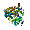
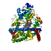
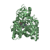
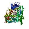
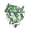
[English] 日本語
Yorodumi
- PDB-2j0d: Crystal structure of human P450 3A4 in complex with erythromycin -

+
Open data


ID or keywords:

Loading...

-
Basic information

Entry
Database: PDB / ID: 2j0d
TitleCrystal structure of human P450 3A4 in complex with erythromycin
ComponentsCYTOCHROME P450 3A4CYP3A4
KeywordsOXIDOREDUCTASE / POLYMORPHISM / ERYTHROMYCIN / MONOOXYGENASE / METAL-BINDING / TRANSMEMBRANE / ENDOPLASMIC RETICULUM / DRUG METABOLIZING ENZYME / P450 / NADP / IRON / HEME / CYP3A4 / MEMBRANE / MICROSOME
Function / homology
Function and homology information


quinine 3-monooxygenase / 1,8-cineole 2-exo-monooxygenase / albendazole monooxygenase (sulfoxide-forming) / quinine 3-monooxygenase activity / 1,8-cineole 2-exo-monooxygenase activity / 1-alpha,25-dihydroxyvitamin D3 23-hydroxylase activity / vitamin D3 25-hydroxylase activity / testosterone 6-beta-hydroxylase activity / vitamin D 24-hydroxylase activity / vitamin D catabolic process ...quinine 3-monooxygenase / 1,8-cineole 2-exo-monooxygenase / albendazole monooxygenase (sulfoxide-forming) / quinine 3-monooxygenase activity / 1,8-cineole 2-exo-monooxygenase activity / 1-alpha,25-dihydroxyvitamin D3 23-hydroxylase activity / vitamin D3 25-hydroxylase activity / testosterone 6-beta-hydroxylase activity / vitamin D 24-hydroxylase activity / vitamin D catabolic process / retinoic acid 4-hydroxylase activity / aflatoxin metabolic process / caffeine oxidase activity / estrogen 16-alpha-hydroxylase activity / estrogen 2-hydroxylase activity / lipid hydroxylation / alkaloid catabolic process / anandamide 8,9 epoxidase activity / anandamide 11,12 epoxidase activity / anandamide 14,15 epoxidase activity / : / Aflatoxin activation and detoxification / Biosynthesis of maresin-like SPMs / monoterpenoid metabolic process / vitamin D metabolic process / Atorvastatin ADME / steroid catabolic process / Xenobiotics / oxidative demethylation / steroid hydroxylase activity / Phase I - Functionalization of compounds / long-chain fatty acid biosynthetic process / estrogen metabolic process / retinoic acid metabolic process / retinol metabolic process / Prednisone ADME / unspecific monooxygenase / aromatase activity / Aspirin ADME / steroid metabolic process / androgen metabolic process / xenobiotic catabolic process / cholesterol metabolic process / steroid binding / xenobiotic metabolic process / monooxygenase activity / lipid metabolic process / oxygen binding / oxidoreductase activity / iron ion binding / intracellular membrane-bounded organelle / heme binding / endoplasmic reticulum membrane / enzyme binding / cytoplasm
Similarity search - Function
Cytochrome P450, E-class, group II / Cytochrome P450, E-class, CYP3A / Cytochrome p450 / Cytochrome P450 / Cytochrome P450, conserved site / Cytochrome P450 cysteine heme-iron ligand signature. / Cytochrome P450 / Cytochrome P450 superfamily / Cytochrome P450 / Orthogonal Bundle / Mainly Alpha
Similarity search - Domain/homology
ERYTHROMYCIN A / PROTOPORPHYRIN IX CONTAINING FE / Cytochrome P450 3A4
Similarity search - Component
Biological speciesHOMO SAPIENS (human)
MethodX-RAY DIFFRACTION / SYNCHROTRON / MOLECULAR REPLACEMENT / Resolution: 2.75 Å
AuthorsSjogren, T. / Ekroos, M.
CitationJournal: Proc.Natl.Acad.Sci.USA / Year: 2006
Title: Structural Basis for Ligand Promiscuity in Cytochrome P450 3A4
Authors: Ekroos, M. / Sjogren, T.
History
DepositionAug 2, 2006Deposition site: PDBE / Processing site: PDBE
Revision 1.0Sep 4, 2006Provider: repository / Type: Initial release
Revision 1.1May 8, 2011Group: Version format compliance
Revision 1.2Jul 13, 2011Group: Version format compliance
Revision 1.3May 8, 2019Group: Data collection / Experimental preparation / Other
Category: database_PDB_rev / database_PDB_rev_record ...database_PDB_rev / database_PDB_rev_record / exptl_crystal_grow / pdbx_database_proc / pdbx_database_status
Item: _exptl_crystal_grow.method / _pdbx_database_status.recvd_author_approval
Revision 1.4Dec 13, 2023Group: Data collection / Database references ...Data collection / Database references / Derived calculations / Other / Refinement description
Category: chem_comp_atom / chem_comp_bond ...chem_comp_atom / chem_comp_bond / database_2 / pdbx_database_status / pdbx_initial_refinement_model / struct_conn
Item: _database_2.pdbx_DOI / _database_2.pdbx_database_accession ..._database_2.pdbx_DOI / _database_2.pdbx_database_accession / _pdbx_database_status.status_code_sf / _struct_conn.ptnr1_auth_comp_id / _struct_conn.ptnr1_auth_seq_id / _struct_conn.ptnr1_label_asym_id / _struct_conn.ptnr1_label_atom_id / _struct_conn.ptnr1_label_comp_id / _struct_conn.ptnr1_label_seq_id / _struct_conn.ptnr2_auth_comp_id / _struct_conn.ptnr2_auth_seq_id / _struct_conn.ptnr2_label_asym_id / _struct_conn.ptnr2_label_atom_id / _struct_conn.ptnr2_label_comp_id / _struct_conn.ptnr2_label_seq_id

-
Structure visualization

Structure viewerMolecule:
MolmilJmol/JSmol

Downloads & links

-
Assembly

Deposited unit
A: CYTOCHROME P450 3A4
B: CYTOCHROME P450 3A4
hetero molecules


Theoretical massNumber of molelcules
Total (without water)112,9305
Polymers110,9632
Non-polymers1,9673
Water88349
1
A: CYTOCHROME P450 3A4
hetero molecules


Theoretical massNumber of molelcules
Total (without water)56,8323
Polymers55,4811
Non-polymers1,3502
Water181
TypeNameSymmetry operationNumber
identity operation1_555x,y,z1
MethodPQS
2
B: CYTOCHROME P450 3A4
hetero molecules


Theoretical massNumber of molelcules
Total (without water)56,0982
Polymers55,4811
Non-polymers6161
Water181
TypeNameSymmetry operationNumber
identity operation1_555x,y,z1
MethodPQS
Unit cell
Length a, b, c (Å)67.254, 210.706, 161.246
Angle α, β, γ (deg.)90.00, 90.00, 90.00
Int Tables number20
Space group name H-MC2221

-
Components

#1: Protein CYTOCHROME P450 3A4 / CYP3A4 / QUININE 3-MONOOXYGENASE / CYPIIIA4 / NIFEDIPINE OXIDASE / TAUROCHENODEOXYCHOLATE 6-ALPHA- ...QUININE 3-MONOOXYGENASE / CYPIIIA4 / NIFEDIPINE OXIDASE / TAUROCHENODEOXYCHOLATE 6-ALPHA-HYDROXYLASE / NF-25 / P450-PCN1


Mass: 55481.480 Da / Num. of mol.: 2 / Fragment: SOLUBLE DOMAIN, RESIDUES 24-502
Source method: isolated from a genetically manipulated source
Source: (gene. exp.) HOMO SAPIENS (human) / Organ: LIVER / Production host: ESCHERICHIA COLI (E. coli) / Strain (production host): XL1 BLUE
References: UniProt: P08684, unspecific monooxygenase, EC: 1.14.13.67, EC: 1.14.13.97
#2: Chemical ChemComp-HEM / PROTOPORPHYRIN IX CONTAINING FE / HEME / Heme B


Mass: 616.487 Da / Num. of mol.: 2 / Source method: obtained synthetically / Formula: C34H32FeN4O4
#3: Chemical ChemComp-ERY / ERYTHROMYCIN A / Erythromycin


Mass: 733.927 Da / Num. of mol.: 1 / Source method: obtained synthetically / Formula: C37H67NO13 / Comment: antibiotic*YM
#4: Water ChemComp-HOH / water / Water


Mass: 18.015 Da / Num. of mol.: 49 / Source method: isolated from a natural source / Formula: H2O
Sequence detailsTHE SEQUENCE CONFLICT CONFLICT HAS BEEN DESRCRIBED BY GONZALEZ ET AL., DNA VOL: 7, PAGES 79-86, 1998

-
Experimental details

-
Experiment

ExperimentMethod: X-RAY DIFFRACTION / Number of used crystals: 1

-
Sample preparation

CrystalDensity Matthews: 2.7 Å3/Da / Density % sol: 53 %
Crystal growMethod: vapor diffusion, hanging drop / pH: 7.5
Details: HANGING DROP CRYSTALLISATION USING 1:1 RATIO OF PROTEIN AT 20-27 MG/ML AGAINST 0.1 M HEPES PH 7-7.5, 0.2 M LITHIUM SULFATE,0.8-1.0 M LITHIUM CHLORIDE AND 30% PEG4000

-
Data collection

DiffractionMean temperature: 100 K
Diffraction sourceSource: SYNCHROTRON / Site: ESRF / Beamline: ID14-4 / Wavelength: 0.939
DetectorType: ADSC CCD / Detector: CCD / Date: Jul 19, 2005
RadiationProtocol: SINGLE WAVELENGTH / Monochromatic (M) / Laue (L): M / Scattering type: x-ray
Radiation wavelengthWavelength: 0.939 Å / Relative weight: 1
ReflectionResolution: 2.75→29.8 Å / Num. obs: 30291 / % possible obs: 99.9 % / Observed criterion σ(I): 2 / Redundancy: 7.2 % / Rmerge(I) obs: 0.1 / Net I/σ(I): 16.4
Reflection shellResolution: 2.75→2.82 Å / Redundancy: 7.4 % / Rmerge(I) obs: 0.56 / Mean I/σ(I) obs: 3.9 / % possible all: 100

-
Processing

Software
NameVersionClassification
REFMAC5.2.0019refinement
MOSFLMdata reduction
SCALAdata scaling
MOLREPphasing
RefinementMethod to determine structure: MOLECULAR REPLACEMENT
Starting model: PDB ENTRY 1W0E
Resolution: 2.75→30 Å / Cor.coef. Fo:Fc: 0.911 / Cor.coef. Fo:Fc free: 0.853 / SU B: 15.723 / SU ML: 0.331 / Cross valid method: THROUGHOUT / ESU R Free: 0.455 / Stereochemistry target values: MAXIMUM LIKELIHOOD / Details: HYDROGENS HAVE BEEN ADDED IN THE RIDING POSITIONS.
RfactorNum. reflection% reflectionSelection details
Rfree0.315 1483 5 %RANDOM
Rwork0.238 ---
obs0.242 27971 97.3 %-
Solvent computationIon probe radii: 0.8 Å / Shrinkage radii: 0.8 Å / VDW probe radii: 1.4 Å / Solvent model: BABINET MODEL WITH MASK
Refinement stepCycle: LAST / Resolution: 2.75→30 Å
ProteinNucleic acidLigandSolventTotal
Num. atoms7140 0 137 49 7326
Refine LS restraints
Refine-IDTypeDev idealDev ideal targetNumber
X-RAY DIFFRACTIONr_bond_refined_d0.0110.0237462
X-RAY DIFFRACTIONr_bond_other_d
X-RAY DIFFRACTIONr_angle_refined_deg1.3322.03110123
X-RAY DIFFRACTIONr_angle_other_deg
X-RAY DIFFRACTIONr_dihedral_angle_1_deg5.8355881
X-RAY DIFFRACTIONr_dihedral_angle_2_deg36.89923.775302
X-RAY DIFFRACTIONr_dihedral_angle_3_deg21.009151334
X-RAY DIFFRACTIONr_dihedral_angle_4_deg15.9351541
X-RAY DIFFRACTIONr_chiral_restr0.0890.21128
X-RAY DIFFRACTIONr_gen_planes_refined0.0040.025486
X-RAY DIFFRACTIONr_gen_planes_other
X-RAY DIFFRACTIONr_nbd_refined0.2190.23596
X-RAY DIFFRACTIONr_nbd_other
X-RAY DIFFRACTIONr_nbtor_refined0.3130.25133
X-RAY DIFFRACTIONr_nbtor_other
X-RAY DIFFRACTIONr_xyhbond_nbd_refined0.2250.2252
X-RAY DIFFRACTIONr_xyhbond_nbd_other
X-RAY DIFFRACTIONr_metal_ion_refined
X-RAY DIFFRACTIONr_metal_ion_other
X-RAY DIFFRACTIONr_symmetry_vdw_refined0.2170.249
X-RAY DIFFRACTIONr_symmetry_vdw_other
X-RAY DIFFRACTIONr_symmetry_hbond_refined0.2850.22
X-RAY DIFFRACTIONr_symmetry_hbond_other
X-RAY DIFFRACTIONr_symmetry_metal_ion_refined
X-RAY DIFFRACTIONr_symmetry_metal_ion_other
X-RAY DIFFRACTIONr_mcbond_it0.5331.54581
X-RAY DIFFRACTIONr_mcbond_other
X-RAY DIFFRACTIONr_mcangle_it0.96527294
X-RAY DIFFRACTIONr_mcangle_other
X-RAY DIFFRACTIONr_scbond_it1.14133230
X-RAY DIFFRACTIONr_scbond_other
X-RAY DIFFRACTIONr_scangle_it1.8394.52825
X-RAY DIFFRACTIONr_scangle_other
X-RAY DIFFRACTIONr_long_range_B_refined
X-RAY DIFFRACTIONr_long_range_B_other
X-RAY DIFFRACTIONr_rigid_bond_restr
X-RAY DIFFRACTIONr_sphericity_free
X-RAY DIFFRACTIONr_sphericity_bonded
LS refinement shellResolution: 2.75→2.82 Å / Total num. of bins used: 20
RfactorNum. reflection% reflection
Rfree0.404 94 -
Rwork0.294 1916 -
obs--91.61 %

+
About Yorodumi

-
News

-
Feb 9, 2022. New format data for meta-information of EMDB entries

New format data for meta-information of EMDB entries

  • Version 3 of the EMDB header file is now the official format.
  • The previous official version 1.9 will be removed from the archive.

Related info.:EMDB header

External links:wwPDB to switch to version 3 of the EMDB data model

-
Aug 12, 2020. Covid-19 info

Covid-19 info

URL: https://pdbj.org/emnavi/covid19.php

New page: Covid-19 featured information page in EM Navigator.

Related info.:Covid-19 info / Mar 5, 2020. Novel coronavirus structure data

+
Mar 5, 2020. Novel coronavirus structure data

Novel coronavirus structure data

Related info.:Yorodumi Speices / Aug 12, 2020. Covid-19 info

External links:COVID-19 featured content - PDBj / Molecule of the Month (242):Coronavirus Proteases

+
Jan 31, 2019. EMDB accession codes are about to change! (news from PDBe EMDB page)

EMDB accession codes are about to change! (news from PDBe EMDB page)

  • The allocation of 4 digits for EMDB accession codes will soon come to an end. Whilst these codes will remain in use, new EMDB accession codes will include an additional digit and will expand incrementally as the available range of codes is exhausted. The current 4-digit format prefixed with “EMD-” (i.e. EMD-XXXX) will advance to a 5-digit format (i.e. EMD-XXXXX), and so on. It is currently estimated that the 4-digit codes will be depleted around Spring 2019, at which point the 5-digit format will come into force.
  • The EM Navigator/Yorodumi systems omit the EMD- prefix.

Related info.:Q: What is EMD? / ID/Accession-code notation in Yorodumi/EM Navigator

External links:EMDB Accession Codes are Changing Soon! / Contact to PDBj

+
Jul 12, 2017. Major update of PDB

Major update of PDB

  • wwPDB released updated PDB data conforming to the new PDBx/mmCIF dictionary.
  • This is a major update changing the version number from 4 to 5, and with Remediation, in which all the entries are updated.
  • In this update, many items about electron microscopy experimental information are reorganized (e.g. em_software).
  • Now, EM Navigator and Yorodumi are based on the updated data.

External links:wwPDB Remediation / Enriched Model Files Conforming to OneDep Data Standards Now Available in the PDB FTP Archive

-
Yorodumi

Thousand views of thousand structures

  • Yorodumi is a browser for structure data from EMDB, PDB, SASBDB, etc.
  • This page is also the successor to EM Navigator detail page, and also detail information page/front-end page for Omokage search.
  • The word "yorodu" (or yorozu) is an old Japanese word meaning "ten thousand". "mi" (miru) is to see.

Related info.:EMDB / PDB / SASBDB / Comparison of 3 databanks / Yorodumi Search / Aug 31, 2016. New EM Navigator & Yorodumi / Yorodumi Papers / Jmol/JSmol / Function and homology information / Changes in new EM Navigator and Yorodumi

Read more