type II site-specific deoxyribonuclease activity / DNA restriction-modification system / magnesium ion binding / DNA binding Similarity search - Function
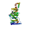
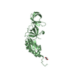
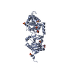
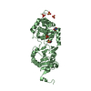
ALTHOUGH SDAI IS A DIMER, NO BIOLOGICALLY RELEVANT DIMERCAN BE CONSTRUCTED IN THE PRESENT STRUCTURE, WHICH ISTHE REASON THIS ENTRY IS MARKED AS MONOMERIC.
-
Components
#1: Protein
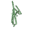
SDAIRESTRICTIONENDONUCLEASE
Mass: 36526.281 Da / Num. of mol.: 2 Source method: isolated from a genetically manipulated source Source: (gene. exp.) STREPTOMYCES DIASTATICUS (bacteria) / Strain: NG7-324 / Plasmid: PAL-SDAIR / Production host: ESCHERICHIA COLI (E. coli) / Strain (production host): ER2267 References: UniProt: C4P954*PLUS, type II site-specific deoxyribonuclease
Monochromator: SI(111) / Protocol: SINGLE WAVELENGTH / Monochromatic (M) / Laue (L): M / Scattering type: x-ray
Radiation wavelength
Wavelength: 0.9755 Å / Relative weight: 1
Reflection
Resolution: 2→51.2 Å / Num. obs: 60505 / % possible obs: 100 % / Observed criterion σ(I): 1.4 / Redundancy: 7.4 % / Biso Wilson estimate: 19.03 Å2 / Rmerge(I) obs: 0.1 / Net I/σ(I): 5.6
Reflection shell
Resolution: 2→2.11 Å / Redundancy: 7.5 % / Rmerge(I) obs: 0.39 / Mean I/σ(I) obs: 1.4 / % possible all: 100
-
Processing
Software
Name
Version
Classification
REFMAC
5.1.24
refinement
MOSFLM
datareduction
SCALA
datascaling
MLPHARE
phasing
Refinement
Method to determine structure: SIRAS / Resolution: 2→30 Å / Cor.coef. Fo:Fc: 0.942 / Cor.coef. Fo:Fc free: 0.925 / SU B: 3.229 / SU ML: 0.09 / Cross valid method: THROUGHOUT / ESU R: 0.157 / ESU R Free: 0.142 / Stereochemistry target values: MAXIMUM LIKELIHOOD Details: HYDROGENS HAVE BEEN ADDED IN THE RIDING POSITIONS.REFINEMENT WAS CONDUCTED WITH CNS, FINAL STEPS OF REFINEMENT WERE MADE WITH REFMAC. RESIDUES 1-4 ARE DISORDERED. SDAI IS A DIMER IN SOLUTION ...Details: HYDROGENS HAVE BEEN ADDED IN THE RIDING POSITIONS.REFINEMENT WAS CONDUCTED WITH CNS, FINAL STEPS OF REFINEMENT WERE MADE WITH REFMAC. RESIDUES 1-4 ARE DISORDERED. SDAI IS A DIMER IN SOLUTION HOWEVER WE COULD NOT IDENTIFY BIOLOGICALLY RELEVANT DIMER IN THE CRYSTAL.
Rfactor
Num. reflection
% reflection
Selection details
Rfree
0.212
6068
10 %
RANDOM
Rwork
0.178
-
-
-
obs
0.182
54406
100 %
-
Solvent computation
Ion probe radii: 0.8 Å / Shrinkage radii: 0.8 Å / VDW probe radii: 1.4 Å / Solvent model: BABINET MODEL WITH MASK
 Movie
Movie Controller


 Open data
 Basic information
 Components
 Keywords
 HYDROLASE /
  Function and homology information

 Authors
 Citation
 Structure visualization
 Downloads & links
 Other downloads
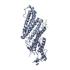

 PDBj
PDBj Assembly
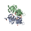

 Mass: 96.063 Da / Num. of mol.: 9 / Source method: obtained synthetically / Formula: SO4
 Mass: 238.305 Da / Num. of mol.: 2 / Source method: obtained synthetically / Formula: C8H18N2O4S / Comment: pH buffer*YM
 Mass: 122.143 Da / Num. of mol.: 2 / Source method: obtained synthetically / Formula: C4H12NO3 / Comment: pH buffer*YM
 Mass: 18.015 Da / Num. of mol.: 470 / Source method: isolated from a natural source / Formula: H2O
 Sample preparation
 / Beamline: ID13 / Wavelength: 0.9755
  Processing