 Movie
Movie Controller

[English] 日本語
 Yorodumi
- PDB-2iwm: precursor mutant Cys1Ser of Penicillin V Acylase from Bacillus sp... -
+ Open data

- Basic information
| Entry | Database: PDB / ID: 2iwm | ||||||
|---|---|---|---|---|---|---|---|
| Title | precursor mutant Cys1Ser of Penicillin V Acylase from Bacillus sphaericus | ||||||
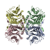
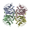
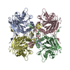
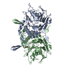
 Components Components | PENICILLIN ACYLASE Penicillin amidase Penicillin amidase | ||||||
 Keywords Keywords | HYDROLASE / ZYMOGEN / PRECURSOR / PENICILLIN / AUTOPROTEOLYSIS / ANTIBIOTIC RESISTANCE | ||||||
| Function / homology |  Function and homology informationpenicillin amidase / penicillin amidase activity / response to antibiotic Function and homology informationpenicillin amidase / penicillin amidase activity / response to antibioticSimilarity search - Function | ||||||
| Biological species |  BACILLUS SPHAERICUS (bacteria) BACILLUS SPHAERICUS (bacteria) | ||||||
| Method | X-RAY DIFFRACTION / SYNCHROTRON / MOLECULAR REPLACEMENT / Resolution: 2.5 Å | ||||||
 Authors Authors | Chandra, P.M. / Dodson, G. / Suresh, C.G. | ||||||
 Citation Citation | Journal: To be Published Title: Precursor Mutant Cys1Ser of Penicillin V Acylase from Bacilus Sphaericus Authors: Chandra, P.M. / Brannigan, J. / Prabhune, A. / Pundle, A. / Turkenburg, J. / Dodson, G. / Suresh, C.G. #1: Journal: Acta Crystallogr.,Sect.F / Year: 2004 Title: Cloning, Preparation and Preliminary Crystallographic Studies of Penicillin V Acylase Autoproteolytic Processing Mutants Authors: Chandra, P.M. / Brannigan, J. / Prabhune, A. / Pundle, A. / Turkenburg, J. / Dodson, G. / Suresh, C.G. | ||||||
| History |
| ||||||
| Remark 700 | SHEET THE SHEET STRUCTURE OF THIS MOLECULE IS BIFURCATED. IN ORDER TO REPRESENT THIS FEATURE IN ... SHEET THE SHEET STRUCTURE OF THIS MOLECULE IS BIFURCATED. IN ORDER TO REPRESENT THIS FEATURE IN THE SHEET RECORDS BELOW, TWO SHEETS ARE DEFINED. |
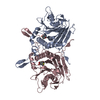
- Structure visualization
| Structure viewer | Molecule: MolmilJmol/JSmol |
|---|
- Downloads & links
-Download
| PDBx/mmCIF format | 2iwm.cif.gz | 264.3 KB | Display | PDBx/mmCIF format |
|---|---|---|---|---|
| PDB format | pdb2iwm.ent.gz | 215.7 KB | Display | PDB format |
| PDBx/mmJSON format | 2iwm.json.gz | Tree view | PDBx/mmJSON format | |
| Others |  Other downloads Other downloads |
-Validation report
| Arichive directory | https://data.pdbj.org/pub/pdb/validation_reports/iw/2iwmftp://data.pdbj.org/pub/pdb/validation_reports/iw/2iwm | HTTPS FTP |
|---|
-Related structure data
| Related structure data | 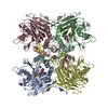 2pvaS S: Starting model for refinement |
|---|---|
| Similar structure data |


-Links
 PDBj
PDBj


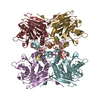
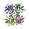
- Assembly
| Deposited unit | 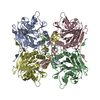
| ||||||||
|---|---|---|---|---|---|---|---|---|---|
| 1 |
| ||||||||
| Unit cell |
|
-Components
| #1: Protein | Penicillin amidase / CYS1SER PRECURSOR MUTANT OF PENICILLIN V ACYLASE / PENICILLIN V AMIDASE / PVA Mass: 37530.535 Da / Num. of mol.: 4 / Mutation: YES Source method: isolated from a genetically manipulated source Details: CYS 4 HAS BEEN MUTATED TO SER BY SITE DIRECTED MUTAGENESIS. RESIDUE CYS1 IN NATIVE PVA IS CYS4 IN THE PRECURSOR STRUCTURE BECAUSE OF ADDITIONAL TRI-PEPTIDE AT N-TERMINUS Source: (gene. exp.) BACILLUS SPHAERICUS (bacteria) / Description: BACILLUS SPHAERICUS NCIM 2478 / Production host: ESCHERICHIA COLI (E. coli) / References: UniProt: P12256, penicillin amidase#2: Water | ChemComp-HOH / | Water Mass: 18.015 Da / Num. of mol.: 323 / Source method: isolated from a natural source / Formula: H2O Mass: 18.015 Da / Num. of mol.: 323 / Source method: isolated from a natural source / Formula: H2OCompound details | ENGINEERED RESIDUE IN CHAIN A, CYS 4 TO SER ENGINEERED RESIDUE IN CHAIN B, CYS 4 TO SER ENGINEERED ...ENGINEERED | |
|---|
-Experimental details
-Experiment
| Experiment | Method: X-RAY DIFFRACTION / Number of used crystals: 1 |
|---|
- Sample preparation
| Crystal | Density Matthews: 3.21 Å3/Da / Density % sol: 61.66 % |
|---|---|
| Crystal grow | pH: 6.4 Details: 700 MICRO LITRE PHOSPHATE BUFFER PH 6.4, 300 MICRO LITRE SATURATED AMMONIUM SULPHATE, 100 MICRO LITRE SUCROSE, 50 MICRO LITRE DMSO |
-Data collection
| Diffraction | Mean temperature: 100 K |
|---|---|
| Diffraction source | Source: SYNCHROTRON / Site: SRS  / Beamline: PX14.2 / Wavelength: 0.978 / Beamline: PX14.2 / Wavelength: 0.978 |
| Detector | Type: ADSC CCD / Detector: CCD / Date: May 5, 2001 |
| Radiation | Protocol: SINGLE WAVELENGTH / Monochromatic (M) / Laue (L): M / Scattering type: x-ray |
| Radiation wavelength | Wavelength: 0.978 Å / Relative weight: 1 |
| Reflection | Resolution: 2.5→20 Å / Num. obs: 62997 / % possible obs: 99.9 % / Observed criterion σ(I): 5 / Redundancy: 3.74 % / Rmerge(I) obs: 0.09 / Net I/σ(I): 16.5 |
- Processing
| Software |
| ||||||||||||||||||||||||||||||||||||||||||||||||||||||||||||||||||||||||||||||||||||||||||||||||||||||||||||||||||||||||||||||||||||||||||||||||||||||||||||||||||||||||||||||||||||||
|---|---|---|---|---|---|---|---|---|---|---|---|---|---|---|---|---|---|---|---|---|---|---|---|---|---|---|---|---|---|---|---|---|---|---|---|---|---|---|---|---|---|---|---|---|---|---|---|---|---|---|---|---|---|---|---|---|---|---|---|---|---|---|---|---|---|---|---|---|---|---|---|---|---|---|---|---|---|---|---|---|---|---|---|---|---|---|---|---|---|---|---|---|---|---|---|---|---|---|---|---|---|---|---|---|---|---|---|---|---|---|---|---|---|---|---|---|---|---|---|---|---|---|---|---|---|---|---|---|---|---|---|---|---|---|---|---|---|---|---|---|---|---|---|---|---|---|---|---|---|---|---|---|---|---|---|---|---|---|---|---|---|---|---|---|---|---|---|---|---|---|---|---|---|---|---|---|---|---|---|---|---|---|---|
| Refinement | Method to determine structure: MOLECULAR REPLACEMENT Starting model: PDB ENTRY 2PVA Resolution: 2.5→100 Å / Cor.coef. Fo:Fc: 0.939 / Cor.coef. Fo:Fc free: 0.897 / SU B: 8.943 / SU ML: 0.2 / Cross valid method: THROUGHOUT / ESU R: 0.407 / ESU R Free: 0.267 / Stereochemistry target values: MAXIMUM LIKELIHOOD / Details: HYDROGENS HAVE BEEN ADDED IN THE RIDING POSITIONS.
| ||||||||||||||||||||||||||||||||||||||||||||||||||||||||||||||||||||||||||||||||||||||||||||||||||||||||||||||||||||||||||||||||||||||||||||||||||||||||||||||||||||||||||||||||||||||
| Solvent computation | Ion probe radii: 0.8 Å / Shrinkage radii: 0.8 Å / VDW probe radii: 1.4 Å / Solvent model: BABINET MODEL WITH MASK | ||||||||||||||||||||||||||||||||||||||||||||||||||||||||||||||||||||||||||||||||||||||||||||||||||||||||||||||||||||||||||||||||||||||||||||||||||||||||||||||||||||||||||||||||||||||
| Displacement parameters | Biso mean: 27.19 Å2
| ||||||||||||||||||||||||||||||||||||||||||||||||||||||||||||||||||||||||||||||||||||||||||||||||||||||||||||||||||||||||||||||||||||||||||||||||||||||||||||||||||||||||||||||||||||||
| Refinement step | Cycle: LAST / Resolution: 2.5→100 Å
| ||||||||||||||||||||||||||||||||||||||||||||||||||||||||||||||||||||||||||||||||||||||||||||||||||||||||||||||||||||||||||||||||||||||||||||||||||||||||||||||||||||||||||||||||||||||
| Refine LS restraints |
|