 Movie
Movie Controller

[English] 日本語
 Yorodumi
- PDB-2itp: Crystal structure of EGFR kinase domain G719S mutation in complex... -
+ Open data

- Basic information
| Entry | Database: PDB / ID: 2itp | ||||||
|---|---|---|---|---|---|---|---|
| Title | Crystal structure of EGFR kinase domain G719S mutation in complex with AEE788 | ||||||
 Components Components | EPIDERMAL GROWTH FACTOR RECEPTOR PRECURSOR | ||||||
 Keywords Keywords |  TRANSFERASE / RECEPTOR / CELL CYCLE / ATP-BINDING / TRANSMEMBRANE / PHOSPHORYLATION / DISEASE MUTATION / POLYMORPHISM / GLYCOPROTEIN / ANTI-ONCOGENE / NUCLEOTIDE-BINDING / ALTERNATIVE SPLICING / AEE788 / EGFR / G719S / KINASE / MEMBRANE / TYROSINE-PROTEIN KINASE / EPIDERMAL GROWTH FACTOR TRANSFERASE / RECEPTOR / CELL CYCLE / ATP-BINDING / TRANSMEMBRANE / PHOSPHORYLATION / DISEASE MUTATION / POLYMORPHISM / GLYCOPROTEIN / ANTI-ONCOGENE / NUCLEOTIDE-BINDING / ALTERNATIVE SPLICING / AEE788 / EGFR / G719S / KINASE / MEMBRANE / TYROSINE-PROTEIN KINASE / EPIDERMAL GROWTH FACTOR | ||||||
| Function / homology |  Function and homology information Function and homology informationresponse to hydroxyisoflavone / multivesicular body, internal vesicle lumen / positive regulation of prolactin secretion / negative regulation of cardiocyte differentiation / positive regulation of protein kinase C activity / diterpenoid metabolic process / Shc-EGFR complex / ovulation cycle / Inhibition of Signaling by Overexpressed EGFR / epidermal growth factor receptor activity ...response to hydroxyisoflavone / multivesicular body, internal vesicle lumen / positive regulation of prolactin secretion / negative regulation of cardiocyte differentiation / positive regulation of protein kinase C activity / diterpenoid metabolic process / Shc-EGFR complex / ovulation cycle / Inhibition of Signaling by Overexpressed EGFR / epidermal growth factor receptor activity / EGFR interacts with phospholipase C-gamma / positive regulation of mucus secretion / response to UV-A / epidermal growth factor binding / PLCG1 events in ERBB2 signaling / tongue development / midgut development / ERBB2-EGFR signaling pathway / hydrogen peroxide metabolic process / PTK6 promotes HIF1A stabilization / digestive tract morphogenesis / regulation of nitric-oxide synthase activity / morphogenesis of an epithelial fold / ERBB2 Activates PTK6 Signaling / intracellular vesicle / Signaling by EGFR / response to cobalamin / transmembrane receptor protein tyrosine kinase activator activity / protein tyrosine kinase activator activity / negative regulation of epidermal growth factor receptor signaling pathway / Signaling by ERBB4 / regulation of phosphatidylinositol 3-kinase/protein kinase B signal transduction / eyelid development in camera-type eye / protein insertion into membrane / cerebral cortex cell migration / ERBB2 Regulates Cell Motility / regulation of JNK cascade / : / PI3K events in ERBB2 signaling / positive regulation of cyclin-dependent protein serine/threonine kinase activity / negative regulation of mitotic cell cycle / hair follicle development / MAP kinase kinase kinase activity / Estrogen-dependent nuclear events downstream of ESR-membrane signaling / embryonic placenta development / positive regulation of bone resorption / positive regulation of G1/S transition of mitotic cell cycle / GAB1 signalosome / positive regulation of nitric oxide mediated signal transduction / salivary gland morphogenesis / peptidyl-tyrosine autophosphorylation / regulation of peptidyl-tyrosine phosphorylation / positive regulation of phosphorylation / positive regulation of glial cell proliferation / positive regulation of vasoconstriction / Signaling by ERBB2 / cellular response to epidermal growth factor stimulus / cellular response to cadmium ion / GRB2 events in EGFR signaling / SHC1 events in EGFR signaling / EGFR Transactivation by Gastrin / positive regulation of DNA repair / GRB2 events in ERBB2 signaling / TFAP2 (AP-2) family regulates transcription of growth factors and their receptors / transmembrane receptor protein tyrosine kinase activity / SHC1 events in ERBB2 signaling / ossification / positive regulation of synaptic transmission, glutamatergic / neurogenesis / cellular response to dexamethasone stimulus / basal plasma membrane / regulation of ERK1 and ERK2 cascade / neuron projection morphogenesis / positive regulation of superoxide anion generation / positive regulation of DNA replication / Signal transduction by L1 / epithelial cell proliferation / cellular response to estradiol stimulus / NOTCH3 Activation and Transmission of Signal to the Nucleus / positive regulation of epithelial cell proliferation / astrocyte activation / liver regeneration / positive regulation of protein localization to plasma membrane / EGFR downregulation / cell surface receptor protein tyrosine kinase signaling pathway / cellular response to amino acid stimulus / positive regulation of smooth muscle cell proliferation / Signaling by ERBB2 TMD/JMD mutants / positive regulation of MAP kinase activity / clathrin-coated endocytic vesicle membrane / lung development / Constitutive Signaling by EGFRvIII / Signaling by ERBB2 ECD mutants / epidermal growth factor receptor signaling pathway / Signaling by ERBB2 KD Mutants / receptor protein-tyrosine kinase / negative regulation of protein catabolic process / Downregulation of ERBB2 signaling / ruffle membrane / kinase bindingSimilarity search - Function | ||||||
| Biological species |  HOMO SAPIENS (human) HOMO SAPIENS (human) | ||||||
| Method | X-RAY DIFFRACTION / SYNCHROTRON / MOLECULAR REPLACEMENT / Resolution: 2.74 Å | ||||||
 Authors Authors | Yun, C.-H. / Boggon, T.J. / Li, Y. / Woo, S. / Greulich, H. / Meyerson, M. / Eck, M.J. | ||||||
 Citation Citation | Journal: Cancer Cell / Year: 2007 Title: Structures of Lung Cancer-Derived Egfr Mutants and Inhibitor Complexes: Mechanism of Activation and Insights Into Differential Inhibitor Sensitivity Authors: Yun, C.-H. / Boggon, T.J. / Li, Y. / Woo, S. / Greulich, H. / Meyerson, M. / Eck, M.J. | ||||||
| History |
|
- Structure visualization
| Structure viewer | Molecule: MolmilJmol/JSmol |
|---|
- Downloads & links
-Download
| PDBx/mmCIF format | 2itp.cif.gz | 78.7 KB | Display | PDBx/mmCIF format |
|---|---|---|---|---|
| PDB format | pdb2itp.ent.gz | 58 KB | Display | PDB format |
| PDBx/mmJSON format | 2itp.json.gz | Tree view | PDBx/mmJSON format | |
| Others |  Other downloads Other downloads |
-Validation report
| Arichive directory | https://data.pdbj.org/pub/pdb/validation_reports/it/2itpftp://data.pdbj.org/pub/pdb/validation_reports/it/2itp | HTTPS FTP |
|---|
-Related structure data
| Related structure data |  2itnC 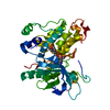 2itoC  2itqC  2ittC  2ituC  2itvC  2itwC  2itxC 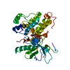 2ityC 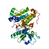 2itzC  2j6mC 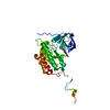 1m17S C: citing same article ( S: Starting model for refinement |
|---|---|
| Similar structure data |


-Links
 PDBj
PDBj


- Assembly
| Deposited unit | 
| ||||||||
|---|---|---|---|---|---|---|---|---|---|
| 1 |
| ||||||||
| Unit cell |
|
-Components
| #1: Protein | Mass: 37334.152 Da / Num. of mol.: 1 / Fragment: KINASE DOMAIN, RESIDUES 696-1022 / Mutation: YES Source method: isolated from a genetically manipulated source Details: EGFR 696-1022 G719S / Source: (gene. exp.) HOMO SAPIENS (human) / Description: EGFR 696-1022 G719S / Plasmid: PACG2T / Cell line (production host): SF9 / Production host:   SPODOPTERA FRUGIPERDA (fall armyworm) SPODOPTERA FRUGIPERDA (fall armyworm)References: UniProt: P00533, receptor protein-tyrosine kinase | ||
|---|---|---|---|
| #2: Chemical | ChemComp-AEE / AEE788  Mass: 440.583 Da / Num. of mol.: 1 / Source method: obtained synthetically / Formula: C27H32N6 / Comment: inhibitor*YM Mass: 440.583 Da / Num. of mol.: 1 / Source method: obtained synthetically / Formula: C27H32N6 / Comment: inhibitor*YM | ||
| #3: Water | ChemComp-HOH / Water Mass: 18.015 Da / Num. of mol.: 65 / Source method: isolated from a natural source / Formula: H2O Mass: 18.015 Da / Num. of mol.: 65 / Source method: isolated from a natural source / Formula: H2O | ||
| Compound details | ENGINEERED| Sequence details | G719S MUTATION | |
-Experimental details
-Experiment
| Experiment | Method: X-RAY DIFFRACTION / Number of used crystals: 1 |
|---|
- Sample preparation
| Crystal | Density Matthews: 3.4 Å3/Da / Density % sol: 64 % |
|---|---|
| Crystal grow | pH: 7.5 / Details: 1.2M KNA TARTRATE, 0.1M HEPES 7.5 |
-Data collection
| Diffraction | Mean temperature: 100 K |
|---|---|
| Diffraction source | Source: SYNCHROTRON / Site: APS  / Beamline: 19-ID / Wavelength: 0.9794 / Beamline: 19-ID / Wavelength: 0.9794 |
| Detector | Type: ADSC CCD / Detector: CCD / Date: Dec 3, 2005 |
| Radiation | Protocol: SINGLE WAVELENGTH / Monochromatic (M) / Laue (L): M / Scattering type: x-ray |
| Radiation wavelength | Wavelength: 0.9794 Å / Relative weight: 1 |
| Reflection | Resolution: 2.74→50 Å / Num. obs: 13390 / % possible obs: 99.9 % / Observed criterion σ(I): -3 / Redundancy: 7.2 % / Rmerge(I) obs: 0.06 / Net I/σ(I): 38.6 |
| Reflection shell | Resolution: 2.74→2.95 Å / Redundancy: 7.4 % / Rmerge(I) obs: 0.37 / Mean I/σ(I) obs: 6.1 / % possible all: 100 |
- Processing
| Software |
| ||||||||||||||||||||||||||||||||||||||||||||||||||||||||||||||||||||||||||||||||||||||||||||||||||||||||||||||||||||||||||||||||||||||||||||||||||||||||||||||||||||||||||||||||||||||
|---|---|---|---|---|---|---|---|---|---|---|---|---|---|---|---|---|---|---|---|---|---|---|---|---|---|---|---|---|---|---|---|---|---|---|---|---|---|---|---|---|---|---|---|---|---|---|---|---|---|---|---|---|---|---|---|---|---|---|---|---|---|---|---|---|---|---|---|---|---|---|---|---|---|---|---|---|---|---|---|---|---|---|---|---|---|---|---|---|---|---|---|---|---|---|---|---|---|---|---|---|---|---|---|---|---|---|---|---|---|---|---|---|---|---|---|---|---|---|---|---|---|---|---|---|---|---|---|---|---|---|---|---|---|---|---|---|---|---|---|---|---|---|---|---|---|---|---|---|---|---|---|---|---|---|---|---|---|---|---|---|---|---|---|---|---|---|---|---|---|---|---|---|---|---|---|---|---|---|---|---|---|---|---|
| Refinement | Method to determine structure: MOLECULAR REPLACEMENT Starting model: PDB ENTRY 1M17 Resolution: 2.74→24.8 Å / Cor.coef. Fo:Fc: 0.945 / Cor.coef. Fo:Fc free: 0.89 / Cross valid method: THROUGHOUT / ESU R: 0.592 / ESU R Free: 0.325 / Stereochemistry target values: MAXIMUM LIKELIHOOD / Details: HYDROGENS HAVE BEEN ADDED IN THE RIDING POSITIONS.
| ||||||||||||||||||||||||||||||||||||||||||||||||||||||||||||||||||||||||||||||||||||||||||||||||||||||||||||||||||||||||||||||||||||||||||||||||||||||||||||||||||||||||||||||||||||||
| Solvent computation | Ion probe radii: 0.8 Å / Shrinkage radii: 0.8 Å / VDW probe radii: 1.2 Å / Solvent model: MASK | ||||||||||||||||||||||||||||||||||||||||||||||||||||||||||||||||||||||||||||||||||||||||||||||||||||||||||||||||||||||||||||||||||||||||||||||||||||||||||||||||||||||||||||||||||||||
| Displacement parameters | Biso mean: 56.3 Å2 | ||||||||||||||||||||||||||||||||||||||||||||||||||||||||||||||||||||||||||||||||||||||||||||||||||||||||||||||||||||||||||||||||||||||||||||||||||||||||||||||||||||||||||||||||||||||
| Refinement step | Cycle: LAST / Resolution: 2.74→24.8 Å
| ||||||||||||||||||||||||||||||||||||||||||||||||||||||||||||||||||||||||||||||||||||||||||||||||||||||||||||||||||||||||||||||||||||||||||||||||||||||||||||||||||||||||||||||||||||||
| Refine LS restraints |
|