 Movie
Movie Controller


+ Open data

- Basic information
| Entry | Database: PDB / ID: 2iqh | ||||||
|---|---|---|---|---|---|---|---|
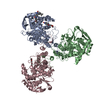
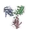
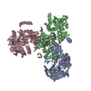
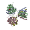
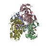
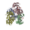
| Title | Influenza A virus nucleoprotein NP at 3.2A resolution | ||||||
 Components Components | Nucleocapsid protein Virus Virus | ||||||
 Keywords Keywords | VIRAL PROTEIN / oligomerization / RNA binding / NLS / polymerase binding | ||||||
| Function / homology |  Function and homology information Function and homology informationhelical viral capsid / viral penetration into host nucleus / viral nucleocapsid / symbiont entry into host cell / ribonucleoprotein complex / host cell nucleus / structural molecule activity / RNA binding / identical protein bindingSimilarity search - Function | ||||||
| Biological species |   Influenza A virus Influenza A virus | ||||||
| Method | X-RAY DIFFRACTION / SYNCHROTRON / AB INITIO PHASING / Resolution: 3.2 Å | ||||||
 Authors Authors | Ye, Q. / Tao, Y.J. | ||||||
 Citation Citation | Journal: Nature / Year: 2006 Title: The mechanism by which influenza A virus nucleoprotein forms oligomers and binds RNA. Authors: Ye, Q. / Krug, R.M. / Tao, Y.J. | ||||||
| History |
|
- Structure visualization
| Structure viewer | Molecule: MolmilJmol/JSmol |
|---|
- Downloads & links
-Download
| PDBx/mmCIF format | 2iqh.cif.gz | 256.4 KB | Display | PDBx/mmCIF format |
|---|---|---|---|---|
| PDB format | pdb2iqh.ent.gz | 209.4 KB | Display | PDB format |
| PDBx/mmJSON format | 2iqh.json.gz | Tree view | PDBx/mmJSON format | |
| Others |  Other downloads Other downloads |
-Validation report
| Arichive directory | https://data.pdbj.org/pub/pdb/validation_reports/iq/2iqhftp://data.pdbj.org/pub/pdb/validation_reports/iq/2iqh | HTTPS FTP |
|---|
-Related structure data
| Similar structure data |
|---|


-Links
 PDBj
PDBj- Assembly
| Deposited unit | 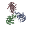
| ||||||||
|---|---|---|---|---|---|---|---|---|---|
| 1 |
| ||||||||
| Unit cell |
|
-Components
| #1: Protein | Virus Mass: 56806.047 Da / Num. of mol.: 3 Source method: isolated from a genetically manipulated source Source: (gene. exp.) Influenza A virus (A/Wilson-Smith/1933(H1N1))Genus: Influenzavirus A / Species: Influenza A virus / Strain: A/Wilson-Smith/1933(H1N1) / Gene: NP / Plasmid: pET28a / Production host:  Escherichia coli (E. coli) / Strain (production host): RosettaTM 2(DE3) SinglesTM / References: UniProt: Q1I2B5, UniProt: Q1K9H2*PLUS Escherichia coli (E. coli) / Strain (production host): RosettaTM 2(DE3) SinglesTM / References: UniProt: Q1I2B5, UniProt: Q1K9H2*PLUS |
|---|
-Experimental details
-Experiment
| Experiment | Method: X-RAY DIFFRACTION / Number of used crystals: 1 |
|---|
- Sample preparation
| Crystal | Density Matthews: 2.36 Å3/Da / Density % sol: 47.9 % |
|---|---|
| Crystal grow | Temperature: 298 K / Method: vapor diffusion, hanging drop / pH: 7.5 Details: 100mM Tris HCl, 3% PEG 8000, 10% Glycerol, 10mM DTT, pH 7.5, VAPOR DIFFUSION, HANGING DROP, temperature 298K |
-Data collection
| Diffraction |
| |||||||||||||||
|---|---|---|---|---|---|---|---|---|---|---|---|---|---|---|---|---|
| Diffraction source |
| |||||||||||||||
| Detector | Type: ADSC QUANTUM 315 / Detector: CCD / Date: Jun 12, 2005 | |||||||||||||||
| Radiation | Protocol: SINGLE WAVELENGTH / Monochromatic (M) / Laue (L): M / Scattering type: x-ray | |||||||||||||||
| Radiation wavelength |
| |||||||||||||||
| Reflection | Resolution: 3.2→30 Å / Num. obs: 26880 / % possible obs: 99.3 % / Observed criterion σ(I): 13.2 / Redundancy: 4.1 % / Rmerge(I) obs: 0.069 / Net I/σ(I): 13.2 | |||||||||||||||
| Reflection shell | Resolution: 3.2→3.31 Å / Redundancy: 4.1 % / Rmerge(I) obs: 0.429 / Mean I/σ(I) obs: 3 / % possible all: 97.9 |

- Processing
| Software |
| ||||||||||||||||||||||||||||||||||||||||||||||||||||||||||||||||||||||||||||||||||||||||||
|---|---|---|---|---|---|---|---|---|---|---|---|---|---|---|---|---|---|---|---|---|---|---|---|---|---|---|---|---|---|---|---|---|---|---|---|---|---|---|---|---|---|---|---|---|---|---|---|---|---|---|---|---|---|---|---|---|---|---|---|---|---|---|---|---|---|---|---|---|---|---|---|---|---|---|---|---|---|---|---|---|---|---|---|---|---|---|---|---|---|---|---|
| Refinement | Method to determine structure: AB INITIO PHASING / Resolution: 3.2→30 Å / Cor.coef. Fo:Fc: 0.885 / Cor.coef. Fo:Fc free: 0.843 / SU B: 30.278 / SU ML: 0.524 / Isotropic thermal model: isotropic / Cross valid method: THROUGHOUT / ESU R Free: 0.653 / Stereochemistry target values: MAXIMUM LIKELIHOOD
| ||||||||||||||||||||||||||||||||||||||||||||||||||||||||||||||||||||||||||||||||||||||||||
| Solvent computation | Ion probe radii: 0.8 Å / Shrinkage radii: 0.8 Å / VDW probe radii: 1.2 Å / Solvent model: BABINET MODEL WITH MASK | ||||||||||||||||||||||||||||||||||||||||||||||||||||||||||||||||||||||||||||||||||||||||||
| Displacement parameters | Biso mean: 74.91 Å2
| ||||||||||||||||||||||||||||||||||||||||||||||||||||||||||||||||||||||||||||||||||||||||||
| Refinement step | Cycle: LAST / Resolution: 3.2→30 Å
| ||||||||||||||||||||||||||||||||||||||||||||||||||||||||||||||||||||||||||||||||||||||||||
| Refine LS restraints |
| ||||||||||||||||||||||||||||||||||||||||||||||||||||||||||||||||||||||||||||||||||||||||||
| LS refinement shell | Resolution: 3.2→3.283 Å / Total num. of bins used: 20
|
