 Movie
Movie Controller


+ Open data

- Basic information
| Entry | Database: PDB / ID: 2io3 | ||||||
|---|---|---|---|---|---|---|---|
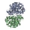
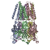
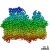
| Title | Crystal structure of human Senp2 in complex with RanGAP1-SUMO-2 | ||||||
 Components Components |
| ||||||
 Keywords Keywords |  PROTEIN BINDING / HYDROLASE / SUMO / Ubiquitin / Senp / Ulp / complex PROTEIN BINDING / HYDROLASE / SUMO / Ubiquitin / Senp / Ulp / complex | ||||||
| Function / homology |  Function and homology information Function and homology informationSUMO-specific endopeptidase activity / cellular response to vasopressin / cytoplasmic periphery of the nuclear pore complex / SUMO ligase complex / SUMOylation of nuclear envelope proteins / SUMO is proteolytically processed / SUMO is conjugated to E1 (UBA2:SAE1) / deSUMOylase activity / protein desumoylation / SUMO is transferred from E1 to E2 (UBE2I, UBC9) ...SUMO-specific endopeptidase activity / cellular response to vasopressin / cytoplasmic periphery of the nuclear pore complex / SUMO ligase complex / SUMOylation of nuclear envelope proteins / SUMO is proteolytically processed / SUMO is conjugated to E1 (UBA2:SAE1) / deSUMOylase activity / protein desumoylation / SUMO is transferred from E1 to E2 (UBE2I, UBC9) / nuclear pore cytoplasmic filaments / Vitamin D (calciferol) metabolism / SUMOylation of SUMOylation proteins / negative regulation of protein export from nucleus / Rev-mediated nuclear export of HIV RNA / regulation of Wnt signaling pathway / activation of GTPase activity / SUMOylation of RNA binding proteins / nuclear export / aggresome / SUMO transferase activity / Postmitotic nuclear pore complex (NPC) reformation / nucleocytoplasmic transport / fat cell differentiation / ubiquitin-like protein ligase binding / SUMOylation of DNA replication proteins / protein sumoylation / SUMOylation of transcription factors / response to axon injury / mRNA transport / Amplification of signal from unattached kinetochores via a MAD2 inhibitory signal / SUMOylation of DNA damage response and repair proteins / nuclear pore / Mitotic Prometaphase / EML4 and NUDC in mitotic spindle formation / axon cytoplasm / negative regulation of protein ubiquitination / Resolution of Sister Chromatid Cohesion / SUMOylation of chromatin organization proteins / GTPase activator activity / SUMOylation of transcription cofactors / positive regulation of protein ubiquitination / RHO GTPases Activate Formins / protein destabilization / SUMOylation of intracellular receptors / mitotic spindle / kinetochore / PML body / small GTPase binding / Wnt signaling pathway / Formation of Incision Complex in GG-NER / protein tag activity / Separation of Sister Chromatids / positive regulation of proteasomal ubiquitin-dependent protein catabolic process / protein transport / nuclear envelope / Processing of DNA double-strand break ends / nuclear membrane / Hydrolases; Acting on peptide bonds (peptidases); Cysteine endopeptidases / cadherin binding / intracellular membrane-bounded organelle / ubiquitin protein ligase binding / dendrite / perinuclear region of cytoplasm / signal transduction / positive regulation of transcription by RNA polymerase II / proteolysis / RNA binding / nucleoplasm / nucleus / cytosol / cytoplasmSimilarity search - Function | ||||||
| Biological species |  Homo sapiens (human) Homo sapiens (human) | ||||||
| Method | X-RAY DIFFRACTION / SYNCHROTRON / MOLECULAR REPLACEMENT / Resolution: 3.2 Å | ||||||
 Authors Authors | Reverter, D. / Lima, C.D. | ||||||
 Citation Citation | Journal: Nat.Struct.Mol.Biol. / Year: 2006 Title: Structural basis for SENP2 protease interactions with SUMO precursors and conjugated substrates. Authors: Reverter, D. / Lima, C.D. | ||||||
| History |
| ||||||
| Remark 300 | BIOMOLECULE: 1 THIS ENTRY CONTAINS THE CRYSTALLOGRAPHIC ASYMMETRIC UNIT WHICH CONSISTS OF 3 ... BIOMOLECULE: 1 THIS ENTRY CONTAINS THE CRYSTALLOGRAPHIC ASYMMETRIC UNIT WHICH CONSISTS OF 3 CHAIN(S). SEE REMARK 350 FOR INFORMATION ON GENERATING THE BIOLOGICAL MOLECULE(S). THERE IS A DOMAIN-SWAPPED DIMER ACROSS CRYSTALLOGRAPHIC TWO-FOLD INVOLVING CHAIN C, FORMED BY SYMMETRY OPERATION 7555 (Y,X,1/3-Z) |
- Structure visualization
| Structure viewer | Molecule: MolmilJmol/JSmol |
|---|
- Downloads & links
-Download
| PDBx/mmCIF format | 2io3.cif.gz | 106.1 KB | Display | PDBx/mmCIF format |
|---|---|---|---|---|
| PDB format | pdb2io3.ent.gz | 80.3 KB | Display | PDB format |
| PDBx/mmJSON format | 2io3.json.gz | Tree view | PDBx/mmJSON format | |
| Others |  Other downloads Other downloads |
-Validation report
| Arichive directory | https://data.pdbj.org/pub/pdb/validation_reports/io/2io3ftp://data.pdbj.org/pub/pdb/validation_reports/io/2io3 | HTTPS FTP |
|---|
-Related structure data
| Related structure data | 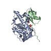 2io0C 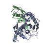 2io1C 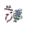 2io2C 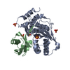 1tgzS S: Starting model for refinement C: citing same article ( |
|---|---|
| Similar structure data |


-Links
 PDBj
PDBj


- Assembly
| Deposited unit | 
| ||||||||
|---|---|---|---|---|---|---|---|---|---|
| 1 | 
| ||||||||
| Unit cell |
|
-Components
| #1: Protein | Mass: 27342.648 Da / Num. of mol.: 1 / Fragment: catalytic domain / Mutation: C548S Source method: isolated from a genetically manipulated source Source: (gene. exp.) Homo sapiens (human) / Gene: SENP2, KIAA1331 / Plasmid: pET28b / Production host:  Escherichia coli (E. coli) Escherichia coli (E. coli)References: UniProt: Q9HC62, Hydrolases; Acting on peptide bonds (peptidases); Cysteine endopeptidases |
|---|---|
| #2: Protein | Mass: 9265.440 Da / Num. of mol.: 1 Source method: isolated from a genetically manipulated source Source: (gene. exp.) Homo sapiens (human) / Gene: SUMO2, SMT3B, SMT3H2 / Plasmid: pET28b / Production host: Escherichia coli (E. coli) / References: UniProt: P61956 |
| #3: Protein | Mass: 18670.543 Da / Num. of mol.: 1 / Fragment: c-terminal domain / Mutation: C573S Source method: isolated from a genetically manipulated source Source: (gene. exp.) Homo sapiens (human) / Gene: RANGAP1 / Plasmid: pET28b / Production host: Escherichia coli (E. coli) / References: UniProt: P46060 |
-Experimental details
-Experiment
| Experiment | Method: X-RAY DIFFRACTION / Number of used crystals: 1 |
|---|
- Sample preparation
| Crystal | Density Matthews: 2.74 Å3/Da / Density % sol: 55.16 % |
|---|---|
| Crystal grow | Temperature: 291 K / Method: vapor diffusion, hanging drop / pH: 8.5 Details: 12% PEG 4000, 0.1M Lithium chloride, 0.1M Tris-HCl, pH 8.5, VAPOR DIFFUSION, HANGING DROP, temperature 291K |
-Data collection
| Diffraction | Mean temperature: 100 K |
|---|---|
| Diffraction source | Source: SYNCHROTRON / Site: NSLS  / Beamline: X29A / Wavelength: 1.1 Å / Beamline: X29A / Wavelength: 1.1 Å |
| Detector | Type: ADSC QUANTUM 315 / Detector: CCD / Date: Jul 14, 2005 |
| Radiation | Protocol: SINGLE WAVELENGTH / Monochromatic (M) / Laue (L): M / Scattering type: x-ray |
| Radiation wavelength | Wavelength: 1.1 Å / Relative weight: 1 |
| Reflection | Resolution: 3.2→50 Å / Num. all: 10581 / Num. obs: 10581 / % possible obs: 98 % / Observed criterion σ(I): 0 / Redundancy: 10.1 % / Rmerge(I) obs: 0.062 / Net I/σ(I): 27.3 |
| Reflection shell | Resolution: 3.2→3.31 Å / Redundancy: 5.4 % / Rmerge(I) obs: 0.248 / Mean I/σ(I) obs: 4.7 / % possible all: 97.3 |
-Phasing
| Phasing MR | Rfactor: 0.49 / Cor.coef. Fo:Fc: 0.665
|
|---|
- Processing
| Software |
| |||||||||||||||||||||||||||||||||||||||||||||||||||||||||
|---|---|---|---|---|---|---|---|---|---|---|---|---|---|---|---|---|---|---|---|---|---|---|---|---|---|---|---|---|---|---|---|---|---|---|---|---|---|---|---|---|---|---|---|---|---|---|---|---|---|---|---|---|---|---|---|---|---|---|
| Refinement | Method to determine structure: MOLECULAR REPLACEMENT Starting model: PDB ENTRY 1TGZ Resolution: 3.2→24.81 Å / Rfactor Rfree error: 0.013 / Data cutoff high absF: 3583109.49 / Data cutoff low absF: 0 / Isotropic thermal model: RESTRAINED / Cross valid method: THROUGHOUT / σ(F): 0
| |||||||||||||||||||||||||||||||||||||||||||||||||||||||||
| Solvent computation | Solvent model: FLAT MODEL / Bsol: 10 Å2 / ksol: 0.190556 e/Å3 | |||||||||||||||||||||||||||||||||||||||||||||||||||||||||
| Displacement parameters | Biso mean: 92.2 Å2
| |||||||||||||||||||||||||||||||||||||||||||||||||||||||||
| Refine analyze |
| |||||||||||||||||||||||||||||||||||||||||||||||||||||||||
| Refinement step | Cycle: LAST / Resolution: 3.2→24.81 Å
| |||||||||||||||||||||||||||||||||||||||||||||||||||||||||
| Refine LS restraints |
| |||||||||||||||||||||||||||||||||||||||||||||||||||||||||
| LS refinement shell | Resolution: 3.2→3.4 Å / Rfactor Rfree error: 0.042 / Total num. of bins used: 6
| |||||||||||||||||||||||||||||||||||||||||||||||||||||||||
| Xplor file |
|
