 Movie
Movie Controller

[English] 日本語
 Yorodumi
- PDB-2inx: Crystal Structure of Ketosteroid Isomerase D40N from Pseudomonas ... -
+ Open data

- Basic information
| Entry | Database: PDB / ID: 2inx | ||||||
|---|---|---|---|---|---|---|---|
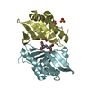
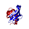
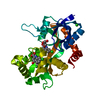
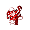
| Title | Crystal Structure of Ketosteroid Isomerase D40N from Pseudomonas putida (pKSI) with bound 2,6-difluorophenol | ||||||
 Components Components | Steroid delta-isomerase | ||||||
 Keywords Keywords | ISOMERASE / KSI / enzyme / active site / charge distribution / hydrogen bond | ||||||
| Function / homology |  Function and homology informationsteroid Delta-isomerase / steroid delta-isomerase activity / steroid metabolic process Function and homology informationsteroid Delta-isomerase / steroid delta-isomerase activity / steroid metabolic processSimilarity search - Function | ||||||
| Biological species |  Pseudomonas putida (bacteria) Pseudomonas putida (bacteria) | ||||||
| Method | X-RAY DIFFRACTION / SYNCHROTRON / MOLECULAR REPLACEMENT / Resolution: 1.5 Å | ||||||
 Authors Authors | Martinez Caaveiro, J.M. / Pybus, B. / Ringe, D. / Petsko, G.A. / Sigala, P. / Kraut, D. / Herschlag, D. | ||||||
 Citation Citation | Journal: J.Am.Chem.Soc. / Year: 2008 Title: Testing geometrical discrimination within an enzyme active site: constrained hydrogen bonding in the ketosteroid isomerase oxyanion hole. Authors: Sigala, P.A. / Kraut, D.A. / Caaveiro, J.M. / Pybus, B. / Ruben, E.A. / Ringe, D. / Petsko, G.A. / Herschlag, D. #1: Journal: PLOS Biol. / Year: 2006Title: Testing Electrostatic Complementarity in Enzyme Catalysis: Hydrogen Bonding in the Ketosteroid Isomerase Oxyanion Hole Authors: Kraut, D. / Sigala, P. / Pybus, B. / Liu, C.W. / Ringe, D. / Petsko, G.A. / Herschlag, D. | ||||||
| History |
|
- Structure visualization
| Structure viewer | Molecule: MolmilJmol/JSmol |
|---|
- Downloads & links
-Download
| PDBx/mmCIF format | 2inx.cif.gz | 67.6 KB | Display | PDBx/mmCIF format |
|---|---|---|---|---|
| PDB format | pdb2inx.ent.gz | 50.1 KB | Display | PDB format |
| PDBx/mmJSON format | 2inx.json.gz | Tree view | PDBx/mmJSON format | |
| Others |  Other downloads Other downloads |
-Validation report
| Arichive directory | https://data.pdbj.org/pub/pdb/validation_reports/in/2inxftp://data.pdbj.org/pub/pdb/validation_reports/in/2inx | HTTPS FTP |
|---|
-Related structure data
| Related structure data |  3cpoC  2b32 S: Starting model for refinement C: citing same article ( |
|---|---|
| Similar structure data |


-Links
 PDBj
PDBj
- Assembly
| Deposited unit | 
| ||||||||
|---|---|---|---|---|---|---|---|---|---|
| 1 | 
| ||||||||
| Unit cell |
| ||||||||
| Components on special symmetry positions |
|
-Components
| #1: Protein | / Delta-5-3-ketosteroid isomerase Mass: 14547.515 Da / Num. of mol.: 1 / Mutation: D40N Source method: isolated from a genetically manipulated source Source: (gene. exp.) Pseudomonas putida (bacteria) / Gene: ksi / Species (production host): Escherichia coli / Production host: Escherichia coli BL21 (bacteria) / Strain (production host): BL21 / References: UniProt: P07445, steroid Delta-isomerase |
|---|---|
| #2: Chemical | ChemComp-FFP /   Mass: 130.092 Da / Num. of mol.: 1 / Source method: obtained synthetically / Formula: C6H4F2O Mass: 130.092 Da / Num. of mol.: 1 / Source method: obtained synthetically / Formula: C6H4F2O |
| #3: Water | ChemComp-HOH / Water Mass: 18.015 Da / Num. of mol.: 72 / Source method: isolated from a natural source / Formula: H2O Mass: 18.015 Da / Num. of mol.: 72 / Source method: isolated from a natural source / Formula: H2O |
-Experimental details
-Experiment
| Experiment | Method: X-RAY DIFFRACTION / Number of used crystals: 1 |
|---|
- Sample preparation
| Crystal | Density Matthews: 2.07 Å3/Da / Density % sol: 40.64 % |
|---|---|
| Crystal grow | Temperature: 298 K / Method: vapor diffusion, hanging drop / pH: 7 Details: Ammonium sulphate 1.4 M, 2-propanol 6.5% protein concentration 25 mg/ml, pH 7.0, VAPOR DIFFUSION, HANGING DROP, temperature 298K |
-Data collection
| Diffraction | Mean temperature: 110 K |
|---|---|
| Diffraction source | Source: SYNCHROTRON / Site: APS  / Beamline: 14-BM-C / Wavelength: 0.9 Å / Beamline: 14-BM-C / Wavelength: 0.9 Å |
| Detector | Type: ADSC QUANTUM 315 / Detector: CCD / Date: Nov 18, 2004 |
| Radiation | Monochromator: Ge 111 / Protocol: SINGLE WAVELENGTH / Monochromatic (M) / Laue (L): M / Scattering type: x-ray |
| Radiation wavelength | Wavelength: 0.9 Å / Relative weight: 1 |
| Reflection | Resolution: 1.5→47.4 Å / Num. all: 19911 / Num. obs: 19274 / % possible obs: 96.8 % / Observed criterion σ(I): -3 / Redundancy: 9 % / Biso Wilson estimate: 24.7 Å2 / Rsym value: 0.079 / Net I/σ(I): 16.1 |
| Reflection shell | Resolution: 1.5→1.55 Å / Redundancy: 8.9 % / Mean I/σ(I) obs: 4.4 / Num. unique all: 1890 / Rsym value: 0.374 / % possible all: 96.7 |
- Processing
| Software |
| |||||||||||||||||||||||||||||||||||||||||||||||||||||||||||||||||||||||||||||||||||||||||||||||||||||||||
|---|---|---|---|---|---|---|---|---|---|---|---|---|---|---|---|---|---|---|---|---|---|---|---|---|---|---|---|---|---|---|---|---|---|---|---|---|---|---|---|---|---|---|---|---|---|---|---|---|---|---|---|---|---|---|---|---|---|---|---|---|---|---|---|---|---|---|---|---|---|---|---|---|---|---|---|---|---|---|---|---|---|---|---|---|---|---|---|---|---|---|---|---|---|---|---|---|---|---|---|---|---|---|---|---|---|---|
| Refinement | Method to determine structure: MOLECULAR REPLACEMENT Starting model: PDB entry 2B32 2b32 Resolution: 1.5→47.4 Å / Cor.coef. Fo:Fc: 0.955 / Cor.coef. Fo:Fc free: 0.928 / SU B: 3.648 / SU ML: 0.062 / Isotropic thermal model: anisotropic / Cross valid method: THROUGHOUT / σ(F): 0 / ESU R: 0.117 / ESU R Free: 0.095 / Stereochemistry target values: MAXIMUM LIKELIHOOD / Details: HYDROGENS HAVE BEEN ADDED IN THE RIDING POSITIONS
| |||||||||||||||||||||||||||||||||||||||||||||||||||||||||||||||||||||||||||||||||||||||||||||||||||||||||
| Solvent computation | Ion probe radii: 0.8 Å / Shrinkage radii: 0.8 Å / VDW probe radii: 1.4 Å / Solvent model: BABINET MODEL WITH MASK | |||||||||||||||||||||||||||||||||||||||||||||||||||||||||||||||||||||||||||||||||||||||||||||||||||||||||
| Displacement parameters | Biso mean: 26.929 Å2
| |||||||||||||||||||||||||||||||||||||||||||||||||||||||||||||||||||||||||||||||||||||||||||||||||||||||||
| Refinement step | Cycle: LAST / Resolution: 1.5→47.4 Å
| |||||||||||||||||||||||||||||||||||||||||||||||||||||||||||||||||||||||||||||||||||||||||||||||||||||||||
| Refine LS restraints |
| |||||||||||||||||||||||||||||||||||||||||||||||||||||||||||||||||||||||||||||||||||||||||||||||||||||||||
| LS refinement shell | Resolution: 1.5→1.537 Å / Total num. of bins used: 20
|
