- PDB-2ims: The X-ray Structure of a Bak Homodimer Reveals an Inhibitory Zinc... -
+
Open data
ID or keywords:
Loading...
-
Basic information
Entry
Database: PDB / ID: 2ims
Title
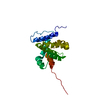
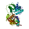
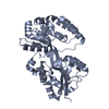
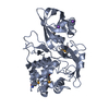
The X-ray Structure of a Bak Homodimer Reveals an Inhibitory Zinc Binding Site
Components
Apoptosis regulator BAK
Keywords
APOPTOSIS / dimer
Function / homology
Function and homology information
Activation and oligomerization of BAK protein / response to mycotoxin / B cell negative selection / BAK complex / BH domain binding / apoptotic process involved in blood vessel morphogenesis / negative regulation of endoplasmic reticulum calcium ion concentration / response to fungus / limb morphogenesis / Release of apoptotic factors from the mitochondria ...Activation and oligomerization of BAK protein / response to mycotoxin / B cell negative selection / BAK complex / BH domain binding / apoptotic process involved in blood vessel morphogenesis / negative regulation of endoplasmic reticulum calcium ion concentration / response to fungus / limb morphogenesis / Release of apoptotic factors from the mitochondria / post-embryonic camera-type eye morphogenesis / endocrine pancreas development / establishment or maintenance of transmembrane electrochemical gradient / positive regulation of mitochondrial outer membrane permeabilization involved in apoptotic signaling pathway / B cell apoptotic process / negative regulation of mitochondrial outer membrane permeabilization involved in apoptotic signaling pathway / activation of cysteine-type endopeptidase activity / endoplasmic reticulum calcium ion homeostasis / positive regulation of endoplasmic reticulum unfolded protein response / regulation of mitochondrial membrane permeability / calcium ion transport into cytosol / response to UV-C / mitochondrial fusion / fibroblast apoptotic process / Bcl-2 family protein complex / myeloid cell homeostasis / positive regulation of calcium ion transport into cytosol / porin activity / thymocyte apoptotic process / pore complex / negative regulation of release of cytochrome c from mitochondria / positive regulation of IRE1-mediated unfolded protein response / positive regulation of release of cytochrome c from mitochondria / positive regulation of proteolysis / vagina development / B cell homeostasis / intrinsic apoptotic signaling pathway in response to endoplasmic reticulum stress / cellular response to unfolded protein / blood vessel remodeling / animal organ regeneration / Pyroptosis / negative regulation of peptidyl-serine phosphorylation / extrinsic apoptotic signaling pathway in absence of ligand / heat shock protein binding / intrinsic apoptotic signaling pathway / release of cytochrome c from mitochondria / regulation of mitochondrial membrane potential / epithelial cell proliferation / establishment of localization in cell / response to gamma radiation / apoptotic signaling pathway / positive regulation of protein-containing complex assembly / response to hydrogen peroxide / response to organic cyclic compound / cellular response to mechanical stimulus / cellular response to UV / intrinsic apoptotic signaling pathway in response to DNA damage / protein-folding chaperone binding / response to ethanol / mitochondrial outer membrane / transmembrane transporter binding / regulation of cell cycle / response to xenobiotic stimulus / positive regulation of apoptotic process / protein heterodimerization activity / negative regulation of cell population proliferation / negative regulation of gene expression / apoptotic process / protein-containing complex binding / endoplasmic reticulum / protein homodimerization activity / mitochondrion / identical protein binding / metal ion binding / cytosol Similarity search - Function
Blc2-like / Apoptosis Regulator Bcl-x / Apoptosis regulator, Bcl-2, BH3 motif, conserved site / Apoptosis regulator, Bcl-2 family BH3 motif signature. / Apoptosis regulator, Bcl-2, BH1 motif, conserved site / Apoptosis regulator, Bcl-2 family BH1 motif signature. / Apoptosis regulator, Bcl-2, BH2 motif, conserved site / Apoptosis regulator, Bcl-2 family BH2 motif signature. / BCL (B-Cell lymphoma); contains BH1, BH2 regions / Bcl-2 family ...Blc2-like / Apoptosis Regulator Bcl-x / Apoptosis regulator, Bcl-2, BH3 motif, conserved site / Apoptosis regulator, Bcl-2 family BH3 motif signature. / Apoptosis regulator, Bcl-2, BH1 motif, conserved site / Apoptosis regulator, Bcl-2 family BH1 motif signature. / Apoptosis regulator, Bcl-2, BH2 motif, conserved site / Apoptosis regulator, Bcl-2 family BH2 motif signature. / BCL (B-Cell lymphoma); contains BH1, BH2 regions / Bcl-2 family / Bcl-2, Bcl-2 homology region 1-3 / Bcl2-like / Apoptosis regulator proteins, Bcl-2 family / BCL2-like apoptosis inhibitors family profile. / Bcl-2-like superfamily / Orthogonal Bundle / Mainly Alpha Similarity search - Domain/homology
Resolution: 1.47→1.52 Å / Redundancy: 2.4 % / Rmerge(I) obs: 0.487 / Mean I/σ(I) obs: 2.17 / Rsym value: 0.348 / % possible all: 86.7
-
Processing
Software
Name
Version
Classification
NB
DENZO
datareduction
SCALEPACK
datascaling
REFMAC
5.1.24
refinement
PDB_EXTRACT
2
dataextraction
ADSC
QUANTUM
datacollection
HKL-2000
datareduction
SOLVE
phasing
Refinement
Method to determine structure: SAD / Resolution: 1.48→52.7 Å / Cor.coef. Fo:Fc: 0.965 / Cor.coef. Fo:Fc free: 0.955 / SU B: 1.305 / SU ML: 0.049 / Cross valid method: THROUGHOUT / ESU R: 0.073 / ESU R Free: 0.073 / Stereochemistry target values: MAXIMUM LIKELIHOOD / Details: HYDROGENS HAVE BEEN ADDED IN THE RIDING POSITIONS
Rfactor
Num. reflection
% reflection
Selection details
Rfree
0.20402
1355
5 %
RANDOM
Rwork
0.17925
-
-
-
obs
0.1805
25678
99.59 %
-
Solvent computation
Ion probe radii: 0.8 Å / Shrinkage radii: 0.8 Å / VDW probe radii: 1.4 Å / Solvent model: BABINET MODEL WITH MASK
Displacement parameters
Biso mean: 19.219 Å2
Baniso -1
Baniso -2
Baniso -3
1-
0.5 Å2
0 Å2
-0.12 Å2
2-
-
0.06 Å2
0 Å2
3-
-
-
-0.66 Å2
Refinement step
Cycle: LAST / Resolution: 1.48→52.7 Å
Protein
Nucleic acid
Ligand
Solvent
Total
Num. atoms
1300
0
1
152
1453
Refine LS restraints
Refine-ID
Type
Dev ideal
Dev ideal target
Number
X-RAY DIFFRACTION
r_bond_refined_d
0.016
0.021
1336
X-RAY DIFFRACTION
r_bond_other_d
0.002
0.02
1180
X-RAY DIFFRACTION
r_angle_refined_deg
1.437
1.922
1812
X-RAY DIFFRACTION
r_angle_other_deg
0.847
3
2724
X-RAY DIFFRACTION
r_dihedral_angle_1_deg
4.713
5
162
X-RAY DIFFRACTION
r_dihedral_angle_2_deg
X-RAY DIFFRACTION
r_dihedral_angle_3_deg
X-RAY DIFFRACTION
r_dihedral_angle_4_deg
X-RAY DIFFRACTION
r_chiral_restr
0.078
0.2
194
X-RAY DIFFRACTION
r_gen_planes_refined
0.007
0.02
1519
X-RAY DIFFRACTION
r_gen_planes_other
0.003
0.02
296
X-RAY DIFFRACTION
r_nbd_refined
0.233
0.2
323
X-RAY DIFFRACTION
r_nbd_other
0.244
0.2
1341
X-RAY DIFFRACTION
r_nbtor_refined
X-RAY DIFFRACTION
r_nbtor_other
0.084
0.2
711
X-RAY DIFFRACTION
r_xyhbond_nbd_refined
0.186
0.2
97
X-RAY DIFFRACTION
r_xyhbond_nbd_other
X-RAY DIFFRACTION
r_metal_ion_refined
0.051
0.2
2
X-RAY DIFFRACTION
r_metal_ion_other
X-RAY DIFFRACTION
r_symmetry_vdw_refined
0.196
0.2
7
X-RAY DIFFRACTION
r_symmetry_vdw_other
0.258
0.2
53
X-RAY DIFFRACTION
r_symmetry_hbond_refined
0.189
0.2
9
X-RAY DIFFRACTION
r_symmetry_hbond_other
X-RAY DIFFRACTION
r_symmetry_metal_ion_refined
X-RAY DIFFRACTION
r_symmetry_metal_ion_other
X-RAY DIFFRACTION
r_mcbond_it
0.968
1.5
808
X-RAY DIFFRACTION
r_mcbond_other
X-RAY DIFFRACTION
r_mcangle_it
1.804
2
1294
X-RAY DIFFRACTION
r_scbond_it
2.968
3
528
X-RAY DIFFRACTION
r_scangle_it
4.75
4.5
518
X-RAY DIFFRACTION
r_rigid_bond_restr
X-RAY DIFFRACTION
r_sphericity_free
X-RAY DIFFRACTION
r_sphericity_bonded
LS refinement shell
Resolution: 1.48→1.513 Å / Total num. of bins used: 20 /
Rfactor
Num. reflection
Rfree
0.289
89
Rwork
0.296
1821
+
About Yorodumi
-
News
-
Feb 9, 2022. New format data for meta-information of EMDB entries
New format data for meta-information of EMDB entries
Version 3 of the EMDB header file is now the official format.
The previous official version 1.9 will be removed from the archive.
In the structure databanks used in Yorodumi, some data are registered as the other names, "COVID-19 virus" and "2019-nCoV". Here are the details of the virus and the list of structure data.
Jan 31, 2019. EMDB accession codes are about to change! (news from PDBe EMDB page)
EMDB accession codes are about to change! (news from PDBe EMDB page)
The allocation of 4 digits for EMDB accession codes will soon come to an end. Whilst these codes will remain in use, new EMDB accession codes will include an additional digit and will expand incrementally as the available range of codes is exhausted. The current 4-digit format prefixed with “EMD-” (i.e. EMD-XXXX) will advance to a 5-digit format (i.e. EMD-XXXXX), and so on. It is currently estimated that the 4-digit codes will be depleted around Spring 2019, at which point the 5-digit format will come into force.
The EM Navigator/Yorodumi systems omit the EMD- prefix.
Related info.:Q: What is EMD? / ID/Accession-code notation in Yorodumi/EM Navigator
Yorodumi is a browser for structure data from EMDB, PDB, SASBDB, etc.
This page is also the successor to EM Navigator detail page, and also detail information page/front-end page for Omokage search.
The word "yorodu" (or yorozu) is an old Japanese word meaning "ten thousand". "mi" (miru) is to see.
Related info.:EMDB / PDB / SASBDB / Comparison of 3 databanks / Yorodumi Search / Aug 31, 2016. New EM Navigator & Yorodumi / Yorodumi Papers / Jmol/JSmol / Function and homology information / Changes in new EM Navigator and Yorodumi
 Movie
Movie Controller

 Yorodumi
 Open data
 Basic information
 Components
 Keywords
 APOPTOSIS / dimer
 Function and homology information
 Authors
 Citation
 Structure visualization
 Downloads & links
 Other downloads
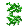


 PDBj
PDBj


 Assembly


 Mass: 65.409 Da / Num. of mol.: 1 / Source method: obtained synthetically / Formula: Zn
 Mass: 18.015 Da / Num. of mol.: 152 / Source method: isolated from a natural source / Formula: H2O
 Sample preparation
 / Beamline: X8C / Wavelength: 1.514 Å
 Processing