 Movie
Movie Controller

[English] 日本語
 Yorodumi
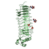
- PDB-2ilr: Crystal structure of human Fanconi Anemia protein E C-terminal domain -
+ Open data

- Basic information
| Entry | Database: PDB / ID: 2ilr | ||||||
|---|---|---|---|---|---|---|---|
| Title | Crystal structure of human Fanconi Anemia protein E C-terminal domain | ||||||
 Components Components | Fanconi anemia group E protein | ||||||
 Keywords Keywords |  ONCOPROTEIN / antiparallel helical hairpin / helical repeat / FANC repeat ONCOPROTEIN / antiparallel helical hairpin / helical repeat / FANC repeat | ||||||
| Function / homology |  Function and homology information Function and homology informationFanconi anaemia nuclear complex / interstrand cross-link repair / Fanconi Anemia Pathway / PKR-mediated signaling / chromosome / centrosome / chromatin / nucleoplasm / nucleus / cytosolSimilarity search - Function | ||||||
| Biological species |  Homo sapiens (human) Homo sapiens (human) | ||||||
| Method | X-RAY DIFFRACTION / SYNCHROTRON / MAD / Resolution: 2 Å | ||||||
 Authors Authors | Pellegrini, L. / Nookala, R.K. | ||||||
 Citation Citation | Journal: Nucleic Acids Res. / Year: 2007 Title: Insights into Fanconi Anaemia from the structure of human FANCE Authors: Nookala, R.K. / Hussain, S. / Pellegrini, L. | ||||||
| History |
|
- Structure visualization
| Structure viewer | Molecule: MolmilJmol/JSmol |
|---|
- Downloads & links
-Download
| PDBx/mmCIF format | 2ilr.cif.gz | 62.7 KB | Display | PDBx/mmCIF format |
|---|---|---|---|---|
| PDB format | pdb2ilr.ent.gz | 46 KB | Display | PDB format |
| PDBx/mmJSON format | 2ilr.json.gz | Tree view | PDBx/mmJSON format | |
| Others |  Other downloads Other downloads |
-Validation report
| Arichive directory | https://data.pdbj.org/pub/pdb/validation_reports/il/2ilrftp://data.pdbj.org/pub/pdb/validation_reports/il/2ilr | HTTPS FTP |
|---|
-Related structure data
| Similar structure data |
|---|


-Links
 PDBj
PDBj- Assembly
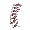
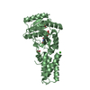
| Deposited unit | 
| ||||||||
|---|---|---|---|---|---|---|---|---|---|
| 1 |
| ||||||||
| Unit cell |
| ||||||||
| Details | The asymmetric unit of the crystal contains one chain of the FANCE protein. This corresponds to the biological unit of the protein. |
-Components
| #1: Protein | Mass: 28986.127 Da / Num. of mol.: 1 / Fragment: C-terminal domain, residues 273-536 / Mutation: C391A Source method: isolated from a genetically manipulated source Source: (gene. exp.) Homo sapiens (human) / Gene: FANCE / Plasmid: pET28a / Species (production host): Escherichia coli / Production host:  Escherichia coli BL21(DE3) (bacteria) / Strain (production host): BL21(DE3) / References: UniProt: Q9HB96 Escherichia coli BL21(DE3) (bacteria) / Strain (production host): BL21(DE3) / References: UniProt: Q9HB96 |
|---|---|
| #2: Water | ChemComp-HOH / Water Mass: 18.015 Da / Num. of mol.: 144 / Source method: isolated from a natural source / Formula: H2O Mass: 18.015 Da / Num. of mol.: 144 / Source method: isolated from a natural source / Formula: H2O |
-Experimental details
-Experiment
| Experiment | Method: X-RAY DIFFRACTION / Number of used crystals: 2 |
|---|
- Sample preparation
| Crystal | Density Matthews: 2.13 Å3/Da / Density % sol: 42.8 % |
|---|---|
| Crystal grow | Temperature: 291 K / Method: vapor diffusion, hanging drop / pH: 8 Details: 0.085M TrisCl, 0.170M Sodium Acetate, 25.5% PEG 4000, 15% glycerol, pH 8.0, VAPOR DIFFUSION, HANGING DROP, temperature 291K |
-Data collection
| Diffraction |
| |||||||||||||||
|---|---|---|---|---|---|---|---|---|---|---|---|---|---|---|---|---|
| Diffraction source |
| |||||||||||||||
| Detector |
| |||||||||||||||
| Radiation |
| |||||||||||||||
| Radiation wavelength |
| |||||||||||||||
| Reflection | Resolution: 2→18.75 Å / Num. all: 17665 / Num. obs: 17665 / % possible obs: 98.9 % / Redundancy: 8.6 % / Biso Wilson estimate: 27.8 Å2 / Rsym value: 0.05 / Net I/σ(I): 8.1 | |||||||||||||||
| Reflection shell | Resolution: 2→2.11 Å / Redundancy: 8.9 % / Num. unique all: 2470 / Rsym value: 0.195 / % possible all: 98.7 |

- Processing
| Software |
| ||||||||||||||||||||||||||||||||||||||||||||||||||||||||||||||||||||||||||||||||||||||||||||||||||||||||||||||||||||||||||||||||||||||||||||||||||||||||||||||||||||||||||
|---|---|---|---|---|---|---|---|---|---|---|---|---|---|---|---|---|---|---|---|---|---|---|---|---|---|---|---|---|---|---|---|---|---|---|---|---|---|---|---|---|---|---|---|---|---|---|---|---|---|---|---|---|---|---|---|---|---|---|---|---|---|---|---|---|---|---|---|---|---|---|---|---|---|---|---|---|---|---|---|---|---|---|---|---|---|---|---|---|---|---|---|---|---|---|---|---|---|---|---|---|---|---|---|---|---|---|---|---|---|---|---|---|---|---|---|---|---|---|---|---|---|---|---|---|---|---|---|---|---|---|---|---|---|---|---|---|---|---|---|---|---|---|---|---|---|---|---|---|---|---|---|---|---|---|---|---|---|---|---|---|---|---|---|---|---|---|---|---|---|---|---|
| Refinement | Method to determine structure: MAD / Resolution: 2→18.75 Å / Cor.coef. Fo:Fc: 0.943 / Cor.coef. Fo:Fc free: 0.927 / SU B: 3.9 / SU ML: 0.111 / Cross valid method: THROUGHOUT / ESU R: 0.201 / ESU R Free: 0.177 / Stereochemistry target values: MAXIMUM LIKELIHOOD / Details: HYDROGENS HAVE BEEN ADDED IN THE RIDING POSITIONS
| ||||||||||||||||||||||||||||||||||||||||||||||||||||||||||||||||||||||||||||||||||||||||||||||||||||||||||||||||||||||||||||||||||||||||||||||||||||||||||||||||||||||||||
| Solvent computation | Ion probe radii: 0.8 Å / Shrinkage radii: 0.8 Å / VDW probe radii: 1.4 Å / Solvent model: BABINET MODEL WITH MASK | ||||||||||||||||||||||||||||||||||||||||||||||||||||||||||||||||||||||||||||||||||||||||||||||||||||||||||||||||||||||||||||||||||||||||||||||||||||||||||||||||||||||||||
| Displacement parameters | Biso mean: 32.895 Å2
| ||||||||||||||||||||||||||||||||||||||||||||||||||||||||||||||||||||||||||||||||||||||||||||||||||||||||||||||||||||||||||||||||||||||||||||||||||||||||||||||||||||||||||
| Refinement step | Cycle: LAST / Resolution: 2→18.75 Å
| ||||||||||||||||||||||||||||||||||||||||||||||||||||||||||||||||||||||||||||||||||||||||||||||||||||||||||||||||||||||||||||||||||||||||||||||||||||||||||||||||||||||||||
| Refine LS restraints |
| ||||||||||||||||||||||||||||||||||||||||||||||||||||||||||||||||||||||||||||||||||||||||||||||||||||||||||||||||||||||||||||||||||||||||||||||||||||||||||||||||||||||||||
| LS refinement shell | Resolution: 2→2.052 Å / Total num. of bins used: 20
|
