 Movie
Movie Controller


+ Open data

- Basic information
| Entry | Database: PDB / ID: 2ijz | ||||||
|---|---|---|---|---|---|---|---|
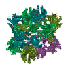
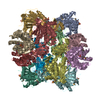
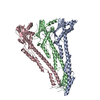
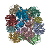
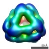
| Title | Crystal structure of aminopeptidase | ||||||
 Components Components | Probable M18-family aminopeptidase 2 | ||||||
 Keywords Keywords |  HYDROLASE / putative aminopeptidase 2 / Pseudomonas aeruginosa / Structural Genomics / PSI / Protein Structure Initiative / New York SGX Research Center for Structural Genomics / NYSGXRC HYDROLASE / putative aminopeptidase 2 / Pseudomonas aeruginosa / Structural Genomics / PSI / Protein Structure Initiative / New York SGX Research Center for Structural Genomics / NYSGXRC | ||||||
| Function / homology |  Function and homology informationHydrolases; Acting on peptide bonds (peptidases); Aminopeptidases / aminopeptidase activity / metallopeptidase activity / proteolysis / zinc ion binding Function and homology informationHydrolases; Acting on peptide bonds (peptidases); Aminopeptidases / aminopeptidase activity / metallopeptidase activity / proteolysis / zinc ion bindingSimilarity search - Function | ||||||
| Biological species |   Pseudomonas aeruginosa (bacteria) Pseudomonas aeruginosa (bacteria) | ||||||
| Method | X-RAY DIFFRACTION / SYNCHROTRON / MOLECULAR REPLACEMENT / Resolution: 3 Å | ||||||
 Authors Authors | Min, T. / Burley, S.K. / Shapiro, L. / New York SGX Research Center for Structural Genomics (NYSGXRC) | ||||||
 Citation Citation | Journal: To be Published Title: Crystal structrue of putative aminopeptidase 2 from Pseudomonas Aeruginosa Authors: Min, T. / Burley, S.K. / Shapiro, L. | ||||||
| History |
|
- Structure visualization
| Structure viewer | Molecule: MolmilJmol/JSmol |
|---|
- Downloads & links
-Download
| PDBx/mmCIF format | 2ijz.cif.gz | 825.9 KB | Display | PDBx/mmCIF format |
|---|---|---|---|---|
| PDB format | pdb2ijz.ent.gz | 667.6 KB | Display | PDB format |
| PDBx/mmJSON format | 2ijz.json.gz | Tree view | PDBx/mmJSON format | |
| Others |  Other downloads Other downloads |
-Validation report
| Arichive directory | https://data.pdbj.org/pub/pdb/validation_reports/ij/2ijzftp://data.pdbj.org/pub/pdb/validation_reports/ij/2ijz | HTTPS FTP |
|---|
-Related structure data
| Related structure data | 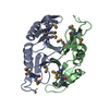 1u6lS S: Starting model for refinement |
|---|---|
| Similar structure data | |
| Other databases |

-Links
 PDBj
PDBj
- Assembly
| Deposited unit | 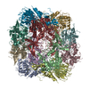
| ||||||||
|---|---|---|---|---|---|---|---|---|---|
| 1 |
| ||||||||
| Unit cell |
|
-Components
| #1: Protein | Mass: 46625.246 Da / Num. of mol.: 12 / Mutation: A154N Source method: isolated from a genetically manipulated source Source: (gene. exp.) Pseudomonas aeruginosa (bacteria) / Gene: apeB / Plasmid: TOPO / Species (production host): Escherichia coli / Production host: Escherichia coli BL21 (bacteria) / Strain (production host): BL21References: UniProt: Q9HYZ3, Hydrolases; Acting on peptide bonds (peptidases); Aminopeptidases#2: Water | ChemComp-HOH / | Water Mass: 18.015 Da / Num. of mol.: 3177 / Source method: isolated from a natural source / Formula: H2O Mass: 18.015 Da / Num. of mol.: 3177 / Source method: isolated from a natural source / Formula: H2O |
|---|
-Experimental details
-Experiment
| Experiment | Method: X-RAY DIFFRACTION / Number of used crystals: 1 |
|---|
- Sample preparation
| Crystal | Density Matthews: 3.09 Å3/Da / Density % sol: 60.19 % |
|---|---|
| Crystal grow | Method: vapor diffusion, hanging drop / pH: 5.6 Details: 0.1M Sodium Citrate, 10% PEG 15K, pH 5.6, VAPOR DIFFUSION, HANGING DROP |
-Data collection
| Diffraction | Mean temperature: 200 K |
|---|---|
| Diffraction source | Source: SYNCHROTRON / Site: NSLS  / Beamline: X29A / Wavelength: 0.9795 Å / Beamline: X29A / Wavelength: 0.9795 Å |
| Detector | Type: ADSC QUANTUM 315 / Detector: CCD / Date: Sep 23, 2006 |
| Radiation | Protocol: SINGLE WAVELENGTH / Monochromatic (M) / Laue (L): M / Scattering type: x-ray |
| Radiation wavelength | Wavelength: 0.9795 Å / Relative weight: 1 |
| Reflection | Resolution: 2.7→20 Å / Num. all: 147805 / Num. obs: 147805 / % possible obs: 87 % / Observed criterion σ(F): 0 / Observed criterion σ(I): 0 / Rmerge(I) obs: 0.106 / Rsym value: 0.106 |
| Reflection shell | Resolution: 2.73→2.9 Å / Redundancy: 1.4 % / Rmerge(I) obs: 0.223 / Num. unique all: 24785 / Rsym value: 0.223 / % possible all: 83.6 |
- Processing
| Software |
| |||||||||||||||||||||||||
|---|---|---|---|---|---|---|---|---|---|---|---|---|---|---|---|---|---|---|---|---|---|---|---|---|---|---|
| Refinement | Method to determine structure: MOLECULAR REPLACEMENT Starting model: pdb entry 1u6l Resolution: 3→20 Å / Rfactor Rfree error: 0.003 / Data cutoff high absF: 212428.56 / Data cutoff low absF: 0 / Isotropic thermal model: GROUP / Cross valid method: THROUGHOUT / σ(F): 2
| |||||||||||||||||||||||||
| Solvent computation | Solvent model: FLAT MODEL / Bsol: 152.868 Å2 / ksol: 0.247676 e/Å3 | |||||||||||||||||||||||||
| Displacement parameters | Biso mean: 10 Å2
| |||||||||||||||||||||||||
| Refine analyze |
| |||||||||||||||||||||||||
| Refinement step | Cycle: LAST / Resolution: 3→20 Å
| |||||||||||||||||||||||||
| Refine LS restraints |
| |||||||||||||||||||||||||
| LS refinement shell | Resolution: 3→3.19 Å / Rfactor Rfree error: 0.008 / Total num. of bins used: 6
| |||||||||||||||||||||||||
| Xplor file |
|
