 Movie
Movie Controller


+ Open data

- Basic information
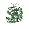
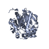
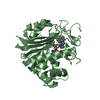
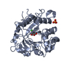
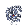
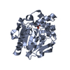
| Entry | Database: PDB / ID: 2iip | ||||||
|---|---|---|---|---|---|---|---|
| Title | Human Nicotinamide N-methyltransferase | ||||||
 Components Components | Nicotinamide N-methyltransferase | ||||||
 Keywords Keywords | TRANSFERASE / mutation / surface mutagenesis / mutant / methyltransferase / Structural Genomics / Structural Genomics Consortium / SGC | ||||||
| Function / homology |  Function and homology informationpyridine N-methyltransferase activity / nicotinamide N-methyltransferase / nicotinamide metabolic process / nicotinamide N-methyltransferase activity / Metabolism of ingested SeMet, Sec, MeSec into H2Se / positive regulation of protein deacetylation / NAD biosynthesis via nicotinamide riboside salvage pathway / Methylation / Nicotinamide salvaging / animal organ regeneration ...pyridine N-methyltransferase activity / nicotinamide N-methyltransferase / nicotinamide metabolic process / nicotinamide N-methyltransferase activity / Metabolism of ingested SeMet, Sec, MeSec into H2Se / positive regulation of protein deacetylation / NAD biosynthesis via nicotinamide riboside salvage pathway / Methylation / Nicotinamide salvaging / animal organ regeneration / positive regulation of gluconeogenesis / response to organonitrogen compound / methylation / response to xenobiotic stimulus / cytosol Function and homology informationpyridine N-methyltransferase activity / nicotinamide N-methyltransferase / nicotinamide metabolic process / nicotinamide N-methyltransferase activity / Metabolism of ingested SeMet, Sec, MeSec into H2Se / positive regulation of protein deacetylation / NAD biosynthesis via nicotinamide riboside salvage pathway / Methylation / Nicotinamide salvaging / animal organ regeneration ...pyridine N-methyltransferase activity / nicotinamide N-methyltransferase / nicotinamide metabolic process / nicotinamide N-methyltransferase activity / Metabolism of ingested SeMet, Sec, MeSec into H2Se / positive regulation of protein deacetylation / NAD biosynthesis via nicotinamide riboside salvage pathway / Methylation / Nicotinamide salvaging / animal organ regeneration / positive regulation of gluconeogenesis / response to organonitrogen compound / methylation / response to xenobiotic stimulus / cytosolSimilarity search - Function | ||||||
| Biological species |  Homo sapiens (human) Homo sapiens (human) | ||||||
| Method | X-RAY DIFFRACTION / MOLECULAR REPLACEMENT / Resolution: 2.05 Å | ||||||
 Authors Authors | Bernstein, G. / Min, J. / Wu, H. / Tempel, W. / Zeng, H. / Loppnau, P. / Avvakumov, G.V. / Wasney, G. / Weigelt, J. / Sundstrom, M. ...Bernstein, G. / Min, J. / Wu, H. / Tempel, W. / Zeng, H. / Loppnau, P. / Avvakumov, G.V. / Wasney, G. / Weigelt, J. / Sundstrom, M. / Arrowsmith, C.H. / Edwards, A.M. / Bochkarev, A. / Plotnikov, A.N. / Structural Genomics Consortium (SGC) | ||||||
 Citation Citation | Journal: To be Published Title: The Crystal Structure of Human Nicotinamide N-methyltransferase in complex with SAH Authors: Bernstein, G. / Min, J. / Wu, H. / Tempel, W. / Zeng, H. / Loppnau, P. / Avvakumov, G.V. / Wasney, G. / Weigelt, J. / Sundstrom, M. / Arrowsmith, C.H. / Edwards, A.M. / Bochkarev, A. / Plotnikov, A.N. | ||||||
| History |
| ||||||
| Remark 999 | The residue Met -18 is a cloning artifact and also an initiating methionine |
- Structure visualization
| Structure viewer | Molecule: MolmilJmol/JSmol |
|---|
- Downloads & links
-Download
| PDBx/mmCIF format | 2iip.cif.gz | 216.1 KB | Display | PDBx/mmCIF format |
|---|---|---|---|---|
| PDB format | pdb2iip.ent.gz | 172.2 KB | Display | PDB format |
| PDBx/mmJSON format | 2iip.json.gz | Tree view | PDBx/mmJSON format | |
| Others |  Other downloads Other downloads |
-Validation report
| Arichive directory | https://data.pdbj.org/pub/pdb/validation_reports/ii/2iipftp://data.pdbj.org/pub/pdb/validation_reports/ii/2iip | HTTPS FTP |
|---|
-Related structure data
| Similar structure data |
|---|


-Links
 PDBj
PDBj- Assembly
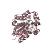
| Deposited unit | 
| ||||||||
|---|---|---|---|---|---|---|---|---|---|
| 1 | 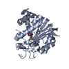
| ||||||||
| 2 | 
| ||||||||
| 3 | 
| ||||||||
| 4 | 
| ||||||||
| Unit cell |
|
-Components
| #1: Protein | Mass: 31466.033 Da / Num. of mol.: 4 / Mutation: K100A, E101A, E103A Source method: isolated from a genetically manipulated source Source: (gene. exp.) Homo sapiens (human) / Gene: NNMT / Production host:  Escherichia coli (E. coli) Escherichia coli (E. coli)References: UniProt: P40261, nicotinamide N-methyltransferase#2: Chemical | ChemComp-SAH / S-Adenosyl-L-homocysteine  Type: L-peptide linking / Mass: 384.411 Da / Num. of mol.: 4 / Source method: obtained synthetically / Formula: C14H20N6O5S Type: L-peptide linking / Mass: 384.411 Da / Num. of mol.: 4 / Source method: obtained synthetically / Formula: C14H20N6O5S#3: Water | ChemComp-HOH / | Water Mass: 18.015 Da / Num. of mol.: 285 / Source method: isolated from a natural source / Formula: H2O Mass: 18.015 Da / Num. of mol.: 285 / Source method: isolated from a natural source / Formula: H2O |
|---|
-Experimental details
-Experiment
| Experiment | Method: X-RAY DIFFRACTION / Number of used crystals: 1 |
|---|
- Sample preparation
| Crystal | Density Matthews: 2.22 Å3/Da / Density % sol: 44.67 % |
|---|---|
| Crystal grow | Temperature: 300 K / Method: vapor diffusion, hanging drop / pH: 7 Details: PEG, pH 7, VAPOR DIFFUSION, HANGING DROP, temperature 300K |
-Data collection
| Diffraction | Mean temperature: 100 K |
|---|---|
| Diffraction source | Source: ROTATING ANODE / Type: RIGAKU / Wavelength: 1.5418 |
| Detector | Type: RIGAKU / Detector: IMAGE PLATE / Date: Sep 20, 2006 |
| Radiation | Monochromator: si / Protocol: SINGLE WAVELENGTH / Monochromatic (M) / Laue (L): M / Scattering type: x-ray |
| Radiation wavelength | Wavelength: 1.5418 Å / Relative weight: 1 |
| Reflection | Resolution: 2.05→29.5 Å / Num. all: 64271 / Num. obs: 64271 / % possible obs: 97.6 % / Observed criterion σ(F): 0 / Observed criterion σ(I): 0 / Redundancy: 2.2 % / Biso Wilson estimate: 40 Å2 / Rmerge(I) obs: 0.091 / Rsym value: 0.091 / Net I/σ(I): 7.7 |
| Reflection shell | Resolution: 2.05→2.12 Å / Redundancy: 2.1 % / Rmerge(I) obs: 0.578 / Mean I/σ(I) obs: 1.4 / Num. unique all: 6482 / Rsym value: 0.578 / % possible all: 93.9 |
- Processing
| Software |
| ||||||||||||||||||||||||||||||||||||||||||||||||||||||||||||||||||||||||||||||||||||||||||
|---|---|---|---|---|---|---|---|---|---|---|---|---|---|---|---|---|---|---|---|---|---|---|---|---|---|---|---|---|---|---|---|---|---|---|---|---|---|---|---|---|---|---|---|---|---|---|---|---|---|---|---|---|---|---|---|---|---|---|---|---|---|---|---|---|---|---|---|---|---|---|---|---|---|---|---|---|---|---|---|---|---|---|---|---|---|---|---|---|---|---|---|
| Refinement | Method to determine structure: MOLECULAR REPLACEMENT / Resolution: 2.05→29.5 Å / Cor.coef. Fo:Fc: 0.939 / Cor.coef. Fo:Fc free: 0.914 / SU B: 5.488 / SU ML: 0.152 / Cross valid method: THROUGHOUT / σ(F): 0 / σ(I): 0 / ESU R: 0.24 / ESU R Free: 0.201 / Stereochemistry target values: MAXIMUM LIKELIHOOD / Details: HYDROGENS HAVE BEEN ADDED IN THE RIDING POSITIONS
| ||||||||||||||||||||||||||||||||||||||||||||||||||||||||||||||||||||||||||||||||||||||||||
| Solvent computation | Ion probe radii: 0.8 Å / Shrinkage radii: 0.8 Å / VDW probe radii: 1.4 Å / Solvent model: MASK | ||||||||||||||||||||||||||||||||||||||||||||||||||||||||||||||||||||||||||||||||||||||||||
| Displacement parameters | Biso mean: 29.364 Å2
| ||||||||||||||||||||||||||||||||||||||||||||||||||||||||||||||||||||||||||||||||||||||||||
| Refinement step | Cycle: LAST / Resolution: 2.05→29.5 Å
| ||||||||||||||||||||||||||||||||||||||||||||||||||||||||||||||||||||||||||||||||||||||||||
| Refine LS restraints |
| ||||||||||||||||||||||||||||||||||||||||||||||||||||||||||||||||||||||||||||||||||||||||||
| LS refinement shell | Resolution: 2.05→2.103 Å / Total num. of bins used: 20
|
