 Movie
Movie Controller


+ Open data

- Basic information
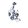
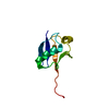
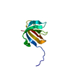
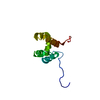
| Entry | Database: PDB / ID: 2ib1 | ||||||
|---|---|---|---|---|---|---|---|
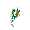
| Title | Solution structure of p45 Death Domain | ||||||
 Components Components | Death domain containing membrane protein NRADD | ||||||
 Keywords Keywords |  APOPTOSIS / p45 / death domain / p75 / Nogo / FADD APOPTOSIS / p45 / death domain / p75 / Nogo / FADD | ||||||
| Function / homology |  Function and homology informationdeath receptor activity / neuron projection membrane / cell body membrane / neurotrophin p75 receptor binding / nerve growth factor binding / nuclear envelope lumen / Rho protein signal transduction / coreceptor activity / lamellipodium / apoptotic process ...death receptor activity / neuron projection membrane / cell body membrane / neurotrophin p75 receptor binding / nerve growth factor binding / nuclear envelope lumen / Rho protein signal transduction / coreceptor activity / lamellipodium / apoptotic process / cell surface / membrane / nucleus / plasma membrane Function and homology informationdeath receptor activity / neuron projection membrane / cell body membrane / neurotrophin p75 receptor binding / nerve growth factor binding / nuclear envelope lumen / Rho protein signal transduction / coreceptor activity / lamellipodium / apoptotic process ...death receptor activity / neuron projection membrane / cell body membrane / neurotrophin p75 receptor binding / nerve growth factor binding / nuclear envelope lumen / Rho protein signal transduction / coreceptor activity / lamellipodium / apoptotic process / cell surface / membrane / nucleus / plasma membraneSimilarity search - Function | ||||||
| Biological species |  Mus musculus (house mouse) Mus musculus (house mouse) | ||||||
| Method | SOLUTION NMR / simulated annealing | ||||||
 Authors Authors | Vilar, M. / Sung, T.C. / Lee, K.F. / Riek, R. | ||||||
 Citation Citation | Journal: To be Published Title: Solution Structure of p45 death domain Authors: Vilar, M. / Sung, T.C. / Lee, K.F. / Riek, R. | ||||||
| History |
|
- Structure visualization
| Structure viewer | Molecule: MolmilJmol/JSmol |
|---|
- Downloads & links
-Download
| PDBx/mmCIF format | 2ib1.cif.gz | 534.9 KB | Display | PDBx/mmCIF format |
|---|---|---|---|---|
| PDB format | pdb2ib1.ent.gz | 447.4 KB | Display | PDB format |
| PDBx/mmJSON format | 2ib1.json.gz | Tree view | PDBx/mmJSON format | |
| Others |  Other downloads Other downloads |
-Validation report
| Arichive directory | https://data.pdbj.org/pub/pdb/validation_reports/ib/2ib1ftp://data.pdbj.org/pub/pdb/validation_reports/ib/2ib1 | HTTPS FTP |
|---|
-Related structure data
| Similar structure data |
|---|


-Links
 PDBj
PDBj


- Assembly
| Deposited unit | 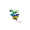
| |||||||||
|---|---|---|---|---|---|---|---|---|---|---|
| 1 |
| |||||||||
| NMR ensembles |
|
-Components
| #1: Protein | Mass: 9975.160 Da / Num. of mol.: 1 / Fragment: residues 138-228 Source method: isolated from a genetically manipulated source Source: (gene. exp.) Mus musculus (house mouse) / Gene: Nradd / Plasmid: pGEX2T / Species (production host): Escherichia coli / Production host:  Escherichia coli BL21 (bacteria) / Strain (production host): BL21 / References: UniProt: Q8CJ26 Escherichia coli BL21 (bacteria) / Strain (production host): BL21 / References: UniProt: Q8CJ26 |
|---|
-Experimental details
-Experiment
| Experiment | Method: SOLUTION NMR | ||||||||||||||||
|---|---|---|---|---|---|---|---|---|---|---|---|---|---|---|---|---|---|
| NMR experiment |
|
- Sample preparation
| Details | Contents: 1-2 mM protein, PBS (Phosphate Buffer Saline) buffer, pH 7.5, 298 K Solvent system: PBS (Phosphate Buffer Saline) buffer, pH 7.5, 298 K |
|---|---|
| Sample conditions | Ionic strength: 137 mM NaCl / pH: 7.5 / Pressure: ambient / Temperature: 298 K |
-NMR measurement
| Radiation | Protocol: SINGLE WAVELENGTH / Monochromatic (M) / Laue (L): M |
|---|---|
| Radiation wavelength | Relative weight: 1 |
| NMR spectrometer | Type: Bruker DMX / Manufacturer: Bruker / Model: DMX / Field strength: 750 MHz |
- Processing
| NMR software |
| ||||||||||||
|---|---|---|---|---|---|---|---|---|---|---|---|---|---|
| Refinement | Method: simulated annealing / Software ordinal: 1 Details: the structures are based on a total of 1319 restraints, 1053 are NOE-derived distance constraints, 266 dihedral angle restraints. | ||||||||||||
| NMR representative | Selection criteria: lowest energy | ||||||||||||
| NMR ensemble | Conformer selection criteria: target function / Conformers calculated total number: 100 / Conformers submitted total number: 20 |
