 Movie
Movie Controller


+ Open data

- Basic information
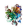
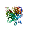
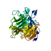
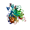
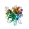
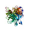
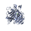
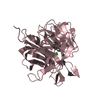
| Entry | Database: PDB / ID: 2iaq | ||||||
|---|---|---|---|---|---|---|---|
| Title | Crystal structure of squid ganglion DFPase S271A mutant | ||||||
 Components Components | Diisopropylfluorophosphatase | ||||||
 Keywords Keywords |  HYDROLASE / phosphotriesterase / beta-propeller / calcium-binding site HYDROLASE / phosphotriesterase / beta-propeller / calcium-binding site | ||||||
| Function / homology |  Function and homology informationdiisopropyl-fluorophosphatase / diisopropyl-fluorophosphatase activity / calcium ion binding Function and homology informationdiisopropyl-fluorophosphatase / diisopropyl-fluorophosphatase activity / calcium ion bindingSimilarity search - Function | ||||||
| Biological species |  Loligo vulgaris (squid) Loligo vulgaris (squid) | ||||||
| Method | X-RAY DIFFRACTION / MOLECULAR REPLACEMENT / Resolution: 2.1 Å | ||||||
 Authors Authors | Scharff, E.I. / Koepke, J. / Fritzsch, G. / Luecke, C. / Rueterjans, H. | ||||||
 Citation Citation | Journal: Structure / Year: 2001 Title: Crystal structure of diisopropylfluorophosphatase from Loligo vulgaris Authors: Scharff, E.I. / Koepke, J. / Fritzsch, G. / Luecke, C. / Rueterjans, H. #1: Journal: Acta Crystallogr.,Sect.D / Year: 2001Title: Crystallization and preliminary X-ray crystallographic analysis of DFPase from Loligo vulgaris Authors: Scharff, E.I. / Luecke, C. / Fritzsch, G. / Koepke, J. / Hartleib, J. / Dierl, S. / Rueterjans, H. #2: Journal: Acta Crystallogr.,Sect.D / Year: 2003Title: Statistical analysis of crystallographic data obtained from squid ganglion DFPase at 0.85 A resolution Authors: Koepke, J. / Scharff, E.I. / Luecke, C. / Rueterjans, H. / Fritzsch, G. #3: Journal: Biochemistry / Year: 2005Title: Mutational and structural studies of the diisopropylfluorophosphatase from Loligo vulgaris shed new light on the catalytic mechanism of the enzyme Authors: Katsemi, V. / Luecke, C. / Koepke, J. / Loehr, F. / Maurer, S. / Fritzsch, G. / Rueterjans, H. | ||||||
| History |
|
- Structure visualization
| Structure viewer | Molecule: MolmilJmol/JSmol |
|---|
- Downloads & links
-Download
| PDBx/mmCIF format | 2iaq.cif.gz | 83.9 KB | Display | PDBx/mmCIF format |
|---|---|---|---|---|
| PDB format | pdb2iaq.ent.gz | 61.1 KB | Display | PDB format |
| PDBx/mmJSON format | 2iaq.json.gz | Tree view | PDBx/mmJSON format | |
| Others |  Other downloads Other downloads |
-Validation report
| Arichive directory | https://data.pdbj.org/pub/pdb/validation_reports/ia/2iaqftp://data.pdbj.org/pub/pdb/validation_reports/ia/2iaq | HTTPS FTP |
|---|
-Related structure data
| Related structure data |  1e1aSC  2iaoC 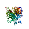 2iapC 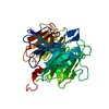 2iarC 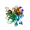 2iasC 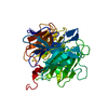 2iatC  2iauC S: Starting model for refinement C: citing same article ( |
|---|---|
| Similar structure data |


-Links
 PDBj
PDBj


- Assembly
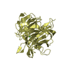
| Deposited unit | 
| ||||||||
|---|---|---|---|---|---|---|---|---|---|
| 1 |
| ||||||||
| Unit cell |
|
-Components
| #1: Protein | Mass: 34844.402 Da / Num. of mol.: 1 / Mutation: S271A Source method: isolated from a genetically manipulated source Details: Phosphotriesterase / Source: (gene. exp.) Loligo vulgaris (squid) / Organ: head ganglion / Plasmid: PKKHISND / Species (production host): Escherichia coli / Production host:  Escherichia coli BL21 (bacteria) / Strain (production host): BL21 / References: UniProt: Q7SIG4, diisopropyl-fluorophosphatase Escherichia coli BL21 (bacteria) / Strain (production host): BL21 / References: UniProt: Q7SIG4, diisopropyl-fluorophosphatase | ||
|---|---|---|---|
| #2: Chemical |   Mass: 40.078 Da / Num. of mol.: 2 / Source method: obtained synthetically / Formula: Ca Mass: 40.078 Da / Num. of mol.: 2 / Source method: obtained synthetically / Formula: Ca#3: Water | ChemComp-HOH / | Water Mass: 18.015 Da / Num. of mol.: 357 / Source method: isolated from a natural source / Formula: H2O Mass: 18.015 Da / Num. of mol.: 357 / Source method: isolated from a natural source / Formula: H2O |
-Experimental details
-Experiment
| Experiment | Method: X-RAY DIFFRACTION / Number of used crystals: 1 |
|---|
- Sample preparation
| Crystal | Density Matthews: 2.17 Å3/Da / Density % sol: 43.25 % |
|---|---|
| Crystal grow | Temperature: 289 K / Method: vapor diffusion, hanging drop / pH: 6.5 Details: 12% PEG 6000, 0.1 M MES, pH 6.5, VAPOR DIFFUSION, HANGING DROP, temperature 289K |
-Data collection
| Diffraction | Mean temperature: 287 K |
|---|---|
| Diffraction source | Source: ROTATING ANODE / Type: RIGAKU / Wavelength: 1.5418 Å |
| Detector | Type: RIGAKU / Detector: IMAGE PLATE / Date: Mar 27, 2000 |
| Radiation | Monochromator: Yale mirrors / Protocol: SINGLE WAVELENGTH / Monochromatic (M) / Laue (L): M / Scattering type: x-ray |
| Radiation wavelength | Wavelength: 1.5418 Å / Relative weight: 1 |
| Reflection | Resolution: 2.1→90 Å / Num. obs: 18311 |
- Processing
| Software |
| ||||||||||||||||||
|---|---|---|---|---|---|---|---|---|---|---|---|---|---|---|---|---|---|---|---|
| Refinement | Method to determine structure: MOLECULAR REPLACEMENT Starting model: PDB entry 1E1A Resolution: 2.1→90 Å / Cross valid method: THROUGHOUT / σ(F): 0 / Stereochemistry target values: Engh & Huber
| ||||||||||||||||||
| Refinement step | Cycle: LAST / Resolution: 2.1→90 Å
|
