 Movie
Movie Controller

[English] 日本語
 Yorodumi
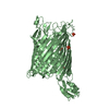
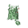
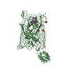
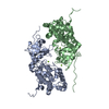
- PDB-2iah: Crystal structure of the ferripyoverdine receptor of the outer me... -
+ Open data

- Basic information
| Entry | Database: PDB / ID: 2iah | ||||||
|---|---|---|---|---|---|---|---|
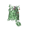
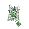
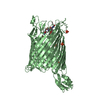
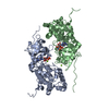
| Title | Crystal structure of the ferripyoverdine receptor of the outer membrane of Pseudomonas aeruginosa bound to ferripyoverdine. | ||||||
 Components Components |
| ||||||
 Keywords Keywords |  MEMBRANE PROTEIN / PYOVERDINE / FPVA / TONB BOX / SIDEROPHORE / CELL MEMBRANE / ION TRANSPORT / TONB DEPENDENT RECEPTOR MEMBRANE PROTEIN / PYOVERDINE / FPVA / TONB BOX / SIDEROPHORE / CELL MEMBRANE / ION TRANSPORT / TONB DEPENDENT RECEPTOR | ||||||
| Function / homology |  Function and homology information Function and homology informationpyoverdine biosynthetic process / siderophore uptake transmembrane transporter activity / cell outer membrane / signaling receptor activity / membraneSimilarity search - Function | ||||||
| Biological species |   Pseudomonas aeruginosa (bacteria) Pseudomonas aeruginosa (bacteria) | ||||||
| Method | X-RAY DIFFRACTION / SYNCHROTRON / MOLECULAR REPLACEMENT / Resolution: 2.73 Å | ||||||
 Authors Authors | Wirth, C. / Pattus, F. / Cobessi, D. | ||||||
 Citation Citation | Journal: J.Mol.Biol. / Year: 2007 Title: From the periplasmic signaling domain to the extracellular face of an outer membrane signal transducer of Pseudomonas aeruginosa: crystal structure of the ferric pyoverdine outer membrane receptor. Authors: Wirth, C. / Meyer-Klaucke, W. / Pattus, F. / Cobessi, D. | ||||||
| History |
|
- Structure visualization
| Structure viewer | Molecule: MolmilJmol/JSmol |
|---|
- Downloads & links
-Download
| PDBx/mmCIF format | 2iah.cif.gz | 164.1 KB | Display | PDBx/mmCIF format |
|---|---|---|---|---|
| PDB format | pdb2iah.ent.gz | 125.8 KB | Display | PDB format |
| PDBx/mmJSON format | 2iah.json.gz | Tree view | PDBx/mmJSON format | |
| Others |  Other downloads Other downloads |
-Validation report
| Arichive directory | https://data.pdbj.org/pub/pdb/validation_reports/ia/2iahftp://data.pdbj.org/pub/pdb/validation_reports/ia/2iah | HTTPS FTP |
|---|
-Related structure data
| Related structure data | 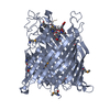 1xkhS S: Starting model for refinement |
|---|---|
| Similar structure data |


-Links
 PDBj
PDBj
- Assembly
| Deposited unit | 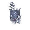
| ||||||||
|---|---|---|---|---|---|---|---|---|---|
| 1 |
| ||||||||
| Unit cell |
|
-Components
| #1: Protein | Mass: 86556.086 Da / Num. of mol.: 1 Source method: isolated from a genetically manipulated source Source: (gene. exp.) Pseudomonas aeruginosa (bacteria) / Gene: fpva / Plasmid: pPVR2 / Production host: Pseudomonas aeruginosa (bacteria) / References: UniProt: P48632 |
|---|---|
| #2: Protein/peptide | 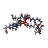  Type: PolypeptidePeptide / Class: Metal transport / Mass: 997.062 Da / Num. of mol.: 1 / Source method: obtained synthetically Type: PolypeptidePeptide / Class: Metal transport / Mass: 997.062 Da / Num. of mol.: 1 / Source method: obtained syntheticallyDetails: IN IRON-DEFICIENT CONDITIONS, PSEUDOMONAS AERUGINOSA SECRETES A MAJOR FLUORESCENT SIDEROPHORE NAMED PYOVERDIN (PVD), WHICH AFTER CHELATING IRON(III) IS TRANSPORTED BACK INTO THE CELL VIA ITS ...Details: IN IRON-DEFICIENT CONDITIONS, PSEUDOMONAS AERUGINOSA SECRETES A MAJOR FLUORESCENT SIDEROPHORE NAMED PYOVERDIN (PVD), WHICH AFTER CHELATING IRON(III) IS TRANSPORTED BACK INTO THE CELL VIA ITS OUTER MEMBRANE RECEPTOR FPVA. FPVA IS A TONB-DEPENDENT TRANSPORT PROTEIN AND HAS THE ABILITY TO BIND PVD IN ITS APO- OR IRON-LOADED FORM. Source: (synth.) Pseudomonas aeruginosa (bacteria) / References: NOR: NOR00190, PYOVERDIN C-E Fe Complex |
| #3: Chemical | ChemComp-SO4 / Sulfate  Mass: 96.063 Da / Num. of mol.: 1 / Source method: obtained synthetically / Formula: SO4 Mass: 96.063 Da / Num. of mol.: 1 / Source method: obtained synthetically / Formula: SO4 |
| #4: Chemical | ChemComp-PVE / (  Type: PolypeptidePeptide / Class: Metal transport / Mass: 376.341 Da / Num. of mol.: 1 / Source method: obtained synthetically / Formula: C17H18N3O7 Type: PolypeptidePeptide / Class: Metal transport / Mass: 376.341 Da / Num. of mol.: 1 / Source method: obtained synthetically / Formula: C17H18N3O7Details: IN IRON-DEFICIENT CONDITIONS, PSEUDOMONAS AERUGINOSA SECRETES A MAJOR FLUORESCENT SIDEROPHORE NAMED PYOVERDIN (PVD), WHICH AFTER CHELATING IRON(III) IS TRANSPORTED BACK INTO THE CELL VIA ITS ...Details: IN IRON-DEFICIENT CONDITIONS, PSEUDOMONAS AERUGINOSA SECRETES A MAJOR FLUORESCENT SIDEROPHORE NAMED PYOVERDIN (PVD), WHICH AFTER CHELATING IRON(III) IS TRANSPORTED BACK INTO THE CELL VIA ITS OUTER MEMBRANE RECEPTOR FPVA. FPVA IS A TONB-DEPENDENT TRANSPORT PROTEIN AND HAS THE ABILITY TO BIND PVD IN ITS APO- OR IRON-LOADED FORM. References: PYOVERDIN C-E Fe Complex |
| #5: Chemical | ChemComp-FE / Iron  Type: PolypeptidePeptide / Class: Metal transport / Mass: 55.845 Da / Num. of mol.: 1 / Source method: obtained synthetically / Formula: Fe Type: PolypeptidePeptide / Class: Metal transport / Mass: 55.845 Da / Num. of mol.: 1 / Source method: obtained synthetically / Formula: FeDetails: IN IRON-DEFICIENT CONDITIONS, PSEUDOMONAS AERUGINOSA SECRETES A MAJOR FLUORESCENT SIDEROPHORE NAMED PYOVERDIN (PVD), WHICH AFTER CHELATING IRON(III) IS TRANSPORTED BACK INTO THE CELL VIA ITS ...Details: IN IRON-DEFICIENT CONDITIONS, PSEUDOMONAS AERUGINOSA SECRETES A MAJOR FLUORESCENT SIDEROPHORE NAMED PYOVERDIN (PVD), WHICH AFTER CHELATING IRON(III) IS TRANSPORTED BACK INTO THE CELL VIA ITS OUTER MEMBRANE RECEPTOR FPVA. FPVA IS A TONB-DEPENDENT TRANSPORT PROTEIN AND HAS THE ABILITY TO BIND PVD IN ITS APO- OR IRON-LOADED FORM. References: PYOVERDIN C-E Fe Complex |
| Compound details | PYOVERDINES ARE A GROUP OF STRUCTURALLY RELATED SIDEROPHORES PRODUCED BY FLUORESCENT PSEUDOMONAS ...PYOVERDINE |
-Experimental details
-Experiment
| Experiment | Method: X-RAY DIFFRACTION / Number of used crystals: 1 |
|---|
- Sample preparation
| Crystal | Density Matthews: 2.79 Å3/Da / Density % sol: 55.86 % |
|---|---|
| Crystal grow | Temperature: 298 K / Method: vapor diffusion, sitting drop / pH: 4.6 Details: Na Acetate 0.1 M, pH 4.6; 12 mM MgSO4, 12 % PEG 3350. Protein concentration in 0.5 % C8E4: 15 mg/ml, VAPOR DIFFUSION, SITTING DROP, temperature 298K |
-Data collection
| Diffraction | Mean temperature: 100 K |
|---|---|
| Diffraction source | Source: SYNCHROTRON / Site: ESRF  / Beamline: ID29 / Wavelength: 0.97857 Å / Beamline: ID29 / Wavelength: 0.97857 Å |
| Detector | Type: ADSC QUANTUM 210 / Detector: CCD / Date: Sep 5, 2005 |
| Radiation | Protocol: SINGLE WAVELENGTH / Monochromatic (M) / Laue (L): M / Scattering type: x-ray |
| Radiation wavelength | Wavelength: 0.97857 Å / Relative weight: 1 |
| Reflection | Resolution: 2.73→46.96 Å / Num. obs: 24636 / % possible obs: 94.4 % / Observed criterion σ(F): 0 / Observed criterion σ(I): -3 / Redundancy: 2.9 % / Rsym value: 0.045 / Net I/σ(I): 7.3 |
| Reflection shell | Resolution: 2.73→2.88 Å / Redundancy: 2.2 % / Mean I/σ(I) obs: 1.3 / Num. unique all: 2982 / Rsym value: 0.274 / % possible all: 78.3 |
- Processing
| Software |
| ||||||||||||||||||||||||||||||||||||||||||||||||||||||||||||||||||||||||||||||||||||||||||||||||||||
|---|---|---|---|---|---|---|---|---|---|---|---|---|---|---|---|---|---|---|---|---|---|---|---|---|---|---|---|---|---|---|---|---|---|---|---|---|---|---|---|---|---|---|---|---|---|---|---|---|---|---|---|---|---|---|---|---|---|---|---|---|---|---|---|---|---|---|---|---|---|---|---|---|---|---|---|---|---|---|---|---|---|---|---|---|---|---|---|---|---|---|---|---|---|---|---|---|---|---|---|---|---|
| Refinement | Method to determine structure: MOLECULAR REPLACEMENT Starting model: 1XKH Resolution: 2.73→46.96 Å / Cor.coef. Fo:Fc: 0.905 / Cor.coef. Fo:Fc free: 0.879 / SU B: 40.321 / SU ML: 0.369 / TLS residual ADP flag: LIKELY RESIDUAL / Cross valid method: THROUGHOUT / σ(F): 0 / ESU R Free: 0.401 / Stereochemistry target values: MAXIMUM LIKELIHOOD
| ||||||||||||||||||||||||||||||||||||||||||||||||||||||||||||||||||||||||||||||||||||||||||||||||||||
| Solvent computation | Ion probe radii: 0.8 Å / Shrinkage radii: 0.8 Å / VDW probe radii: 1.4 Å / Solvent model: MASK | ||||||||||||||||||||||||||||||||||||||||||||||||||||||||||||||||||||||||||||||||||||||||||||||||||||
| Displacement parameters | Biso mean: 51.148 Å2
| ||||||||||||||||||||||||||||||||||||||||||||||||||||||||||||||||||||||||||||||||||||||||||||||||||||
| Refinement step | Cycle: LAST / Resolution: 2.73→46.96 Å
| ||||||||||||||||||||||||||||||||||||||||||||||||||||||||||||||||||||||||||||||||||||||||||||||||||||
| Refine LS restraints |
| ||||||||||||||||||||||||||||||||||||||||||||||||||||||||||||||||||||||||||||||||||||||||||||||||||||
| LS refinement shell | Resolution: 2.73→2.803 Å / Total num. of bins used: 20
| ||||||||||||||||||||||||||||||||||||||||||||||||||||||||||||||||||||||||||||||||||||||||||||||||||||
| Refinement TLS params. | Method: refined / Refine-ID: X-RAY DIFFRACTION
| ||||||||||||||||||||||||||||||||||||||||||||||||||||||||||||||||||||||||||||||||||||||||||||||||||||
| Refinement TLS group |
|
