 Movie
Movie Controller

[English] 日本語
 Yorodumi
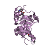
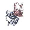
- PDB-2hnv: Crystal Structure of a Dipeptide Complex of the Q58V Mutant of Bo... -
+ Open data

- Basic information
| Entry | Database: PDB / ID: 2hnv | ||||||
|---|---|---|---|---|---|---|---|
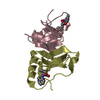
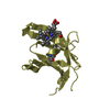
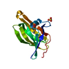
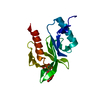
| Title | Crystal Structure of a Dipeptide Complex of the Q58V Mutant of Bovine Neurophysin-I | ||||||
 Components Components | Oxytocin-neurophysin 1 | ||||||
 Keywords Keywords | PEPTIDE BINDING PROTEIN / protein-pepide complex / Q58V mutant / inter-domain loop /  beta sheet / 3 / 10 helix beta sheet / 3 / 10 helix | ||||||
| Function / homology |  Function and homology information Function and homology informationVasopressin-like receptors / oxytocin receptor binding / neurohypophyseal hormone activity / V1A vasopressin receptor binding / neuropeptide hormone activity / G alpha (q) signalling events / secretory granule / response to estrogen / positive regulation of cold-induced thermogenesis / extracellular spaceSimilarity search - Function | ||||||
| Biological species |  Bos taurus (cattle) Bos taurus (cattle) | ||||||
| Method | X-RAY DIFFRACTION / Resolution: 2.5 Å | ||||||
 Authors Authors | Li, X. / Lee, H. / Wu, J. / Breslow, E. | ||||||
 Citation Citation | Journal: Protein Sci. / Year: 2007 Title: Contributions of the interdomain loop, amino terminus, and subunit interface to the ligand-facilitated dimerization of neurophysin: crystal structures and mutation studies of bovine neurophysin-I. Authors: Li, X. / Lee, H. / Wu, J. / Breslow, E. | ||||||
| History |
|
- Structure visualization
| Structure viewer | Molecule: MolmilJmol/JSmol |
|---|
- Downloads & links
-Download
| PDBx/mmCIF format | 2hnv.cif.gz | 85.3 KB | Display | PDBx/mmCIF format |
|---|---|---|---|---|
| PDB format | pdb2hnv.ent.gz | 65.8 KB | Display | PDB format |
| PDBx/mmJSON format | 2hnv.json.gz | Tree view | PDBx/mmJSON format | |
| Others |  Other downloads Other downloads |
-Validation report
| Arichive directory | https://data.pdbj.org/pub/pdb/validation_reports/hn/2hnvftp://data.pdbj.org/pub/pdb/validation_reports/hn/2hnv | HTTPS FTP |
|---|
-Related structure data
| Related structure data | 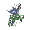 2hnuSC 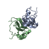 2hnwC S: Starting model for refinement C: citing same article ( |
|---|---|
| Similar structure data |


-Links
 PDBj
PDBj


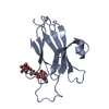
- Assembly
| Deposited unit | 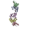
| ||||||||
|---|---|---|---|---|---|---|---|---|---|
| 1 | 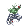
| ||||||||
| 2 | 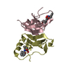
| ||||||||
| 3 | 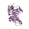
| ||||||||
| Unit cell |
| ||||||||
| Details | The biological unit is a dimer. There are 2.5 biological units per asymmetric unit. The complete dimers are comprised of chains A & B and chains C & D. Chain E is half of a dimer from another asymmetric unit. |
-Components
| #1: Protein | Mass: 8134.256 Da / Num. of mol.: 5 / Fragment: Residues 38-118 / Mutation: Q58V Source method: isolated from a genetically manipulated source Source: (gene. exp.) Bos taurus (cattle) / Gene: OXT / Plasmid: pTHMa30-51 / Production host:  Escherichia coli (E. coli) / Strain (production host): BL21(DE3)p Lys S / References: UniProt: P01175 Escherichia coli (E. coli) / Strain (production host): BL21(DE3)p Lys S / References: UniProt: P01175#2: Chemical | ChemComp-PHE / Phenylalanine  Type: L-peptide linking / Mass: 165.189 Da / Num. of mol.: 5 / Source method: obtained synthetically / Formula: C9H11NO2 Type: L-peptide linking / Mass: 165.189 Da / Num. of mol.: 5 / Source method: obtained synthetically / Formula: C9H11NO2#3: Chemical | ChemComp-TYR / Tyrosine  Type: L-peptide linking / Mass: 181.189 Da / Num. of mol.: 5 / Source method: obtained synthetically / Formula: C9H11NO3 Type: L-peptide linking / Mass: 181.189 Da / Num. of mol.: 5 / Source method: obtained synthetically / Formula: C9H11NO3#4: Water | ChemComp-HOH / | Water Mass: 18.015 Da / Num. of mol.: 29 / Source method: isolated from a natural source / Formula: H2O Mass: 18.015 Da / Num. of mol.: 29 / Source method: isolated from a natural source / Formula: H2O |
|---|
-Experimental details
-Experiment
| Experiment | Method: X-RAY DIFFRACTION / Number of used crystals: 1 |
|---|
- Sample preparation
| Crystal | Density Matthews: 2.65 Å3/Da / Density % sol: 53.67 % |
|---|---|
| Crystal grow | Temperature: 293 K / Method: vapor diffusion, hanging drop / pH: 4 Details: 0.2 M calcium chloride dihydrate, 0.1 M sodium acetate trihydrate, 22% v/v isopropanol, pH 4.0, VAPOR DIFFUSION, HANGING DROP, temperature 293K |
-Data collection
| Diffraction | Mean temperature: 100 K | ||||||||||||||||||||||||||||||||||||||||||||||||||||||||||||||||||
|---|---|---|---|---|---|---|---|---|---|---|---|---|---|---|---|---|---|---|---|---|---|---|---|---|---|---|---|---|---|---|---|---|---|---|---|---|---|---|---|---|---|---|---|---|---|---|---|---|---|---|---|---|---|---|---|---|---|---|---|---|---|---|---|---|---|---|---|
| Diffraction source | Source: ROTATING ANODE / Wavelength: 1.541 Å | ||||||||||||||||||||||||||||||||||||||||||||||||||||||||||||||||||
| Detector | Type: RIGAKU RAXIS IV / Detector: IMAGE PLATE / Date: Jan 1, 2005 / Details: VariMax-HR | ||||||||||||||||||||||||||||||||||||||||||||||||||||||||||||||||||
| Radiation | Protocol: SINGLE WAVELENGTH / Scattering type: x-ray | ||||||||||||||||||||||||||||||||||||||||||||||||||||||||||||||||||
| Radiation wavelength | Wavelength: 1.541 Å / Relative weight: 1 | ||||||||||||||||||||||||||||||||||||||||||||||||||||||||||||||||||
| Reflection | Resolution: 2.5→38.66 Å / Num. obs: 17407 / % possible obs: 99.6 % / Redundancy: 8.2 % / Rmerge(I) obs: 0.074 / Χ2: 1.002 / Net I/σ(I): 15.8 | ||||||||||||||||||||||||||||||||||||||||||||||||||||||||||||||||||
| Reflection shell |
|
- Processing
| Software |
| ||||||||||||||||||||||||||||||||||||
|---|---|---|---|---|---|---|---|---|---|---|---|---|---|---|---|---|---|---|---|---|---|---|---|---|---|---|---|---|---|---|---|---|---|---|---|---|---|
| Refinement | Starting model: PDB ENTRY 2HNU Resolution: 2.5→22.04 Å / Rfactor Rfree error: 0.01 / Data cutoff high absF: 623573.625 / Data cutoff low absF: 0 / Isotropic thermal model: RESTRAINED / Cross valid method: THROUGHOUT / σ(F): 0
| ||||||||||||||||||||||||||||||||||||
| Solvent computation | Solvent model: FLAT MODEL / Bsol: 35.583 Å2 / ksol: 0.335 e/Å3 | ||||||||||||||||||||||||||||||||||||
| Displacement parameters | Biso mean: 56.9 Å2
| ||||||||||||||||||||||||||||||||||||
| Refine analyze |
| ||||||||||||||||||||||||||||||||||||
| Refinement step | Cycle: LAST / Resolution: 2.5→22.04 Å
| ||||||||||||||||||||||||||||||||||||
| Refine LS restraints |
| ||||||||||||||||||||||||||||||||||||
| LS refinement shell | Resolution: 2.5→2.66 Å / Rfactor Rfree error: 0.036 / Total num. of bins used: 6
| ||||||||||||||||||||||||||||||||||||
| Xplor file |
|
