 Movie
Movie Controller

[English] 日本語
 Yorodumi
- PDB-2hj0: Crystal Structure of the Putative Alfa Subunit of Citrate Lyase i... -
+ Open data

- Basic information
| Entry | Database: PDB / ID: 2hj0 | ||||||
|---|---|---|---|---|---|---|---|
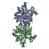
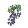
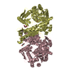
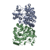
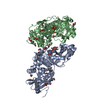
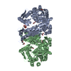
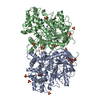
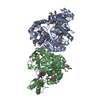
| Title | Crystal Structure of the Putative Alfa Subunit of Citrate Lyase in Complex with Citrate from Streptococcus mutans, Northeast Structural Genomics Target SmR12 . | ||||||
 Components Components | Putative citrate lyase, alfa subunit ATP citrate synthase ATP citrate synthase | ||||||
 Keywords Keywords | LYASE / alpha beta protein. / Structural Genomics / PSI-2 / Protein Structure Initiative / Northeast Structural Genomics Consortium / NESG | ||||||
| Function / homology |  Function and homology informationcitrate CoA-transferase / citrate CoA-transferase activity / ATP-independent citrate lyase complex / citrate (pro-3S)-lyase / citrate (pro-3S)-lyase activity / acetyl-CoA metabolic process Function and homology informationcitrate CoA-transferase / citrate CoA-transferase activity / ATP-independent citrate lyase complex / citrate (pro-3S)-lyase / citrate (pro-3S)-lyase activity / acetyl-CoA metabolic processSimilarity search - Function | ||||||
| Biological species |  Streptococcus mutans (bacteria) Streptococcus mutans (bacteria) | ||||||
| Method | X-RAY DIFFRACTION / SYNCHROTRON / SAD / Resolution: 2.7 Å | ||||||
 Authors Authors | Forouhar, F. / Hussain, M. / Jayaraman, S. / Shastry, R. / Janjua, H. / Cunningham, K. / Ma, L.C. / Xiao, R. / Liu, J. / Baran, M. ...Forouhar, F. / Hussain, M. / Jayaraman, S. / Shastry, R. / Janjua, H. / Cunningham, K. / Ma, L.C. / Xiao, R. / Liu, J. / Baran, M. / Acton, T.B. / Rost, B. / Montelione, G.T. / Tong, L. / Hunt, J.F. / Northeast Structural Genomics Consortium (NESG) | ||||||
 Citation Citation | Journal: To be Published Title: Crystal Structure of the Putative Alfa Subunit of Citrate Lyase in Complex with Citrate from Streptococcus mutans, Northeast Structural Genomics Target SmR12 (CASP Target). Authors: Forouhar, F. / Hussain, M. / Jayaraman, S. / Shastry, R. / Janjua, H. / Cunningham, K. / Ma, L.C. / Xiao, R. / Liu, J. / Baran, M. / Acton, T.B. / Rost, B. / Montelione, G.T. / Tong, L. / Hunt, J.F. | ||||||
| History |
|
- Structure visualization
| Structure viewer | Molecule: MolmilJmol/JSmol |
|---|
- Downloads & links
-Download
| PDBx/mmCIF format | 2hj0.cif.gz | 199.4 KB | Display | PDBx/mmCIF format |
|---|---|---|---|---|
| PDB format | pdb2hj0.ent.gz | 168.2 KB | Display | PDB format |
| PDBx/mmJSON format | 2hj0.json.gz | Tree view | PDBx/mmJSON format | |
| Others |  Other downloads Other downloads |
-Validation report
| Arichive directory | https://data.pdbj.org/pub/pdb/validation_reports/hj/2hj0ftp://data.pdbj.org/pub/pdb/validation_reports/hj/2hj0 | HTTPS FTP |
|---|
-Related structure data
| Similar structure data | |
|---|---|
| Other databases |


-Links
 PDBj
PDBj- Assembly
| Deposited unit | 
| ||||||||
|---|---|---|---|---|---|---|---|---|---|
| 1 |
| ||||||||
| Unit cell |
|
-Components
| #1: Protein | ATP citrate synthase Mass: 57199.773 Da / Num. of mol.: 2 Source method: isolated from a genetically manipulated source Source: (gene. exp.) Streptococcus mutans (bacteria) / Strain: UA159 / Gene: cilA / Plasmid: pET21 / Production host: Escherichia coli (E. coli) / Strain (production host): BL21(DE3)+Magic / References: UniProt: Q8DUC1, citrate (pro-3S)-lyase#2: Chemical | Citric acid  Mass: 192.124 Da / Num. of mol.: 2 / Source method: obtained synthetically / Formula: C6H8O7 Mass: 192.124 Da / Num. of mol.: 2 / Source method: obtained synthetically / Formula: C6H8O7#3: Water | ChemComp-HOH / | Water Mass: 18.015 Da / Num. of mol.: 115 / Source method: isolated from a natural source / Formula: H2O Mass: 18.015 Da / Num. of mol.: 115 / Source method: isolated from a natural source / Formula: H2O |
|---|
-Experimental details
-Experiment
| Experiment | Method: X-RAY DIFFRACTION / Number of used crystals: 1 |
|---|
- Sample preparation
| Crystal | Density Matthews: 2.5 Å3/Da / Density % sol: 50.74 % |
|---|---|
| Crystal grow | Temperature: 293 K / Method: vapor diffusion, sitting drop / pH: 7.5 Details: 100mM HEPES, 10% PEG20k, 5mM citrate, and 5mM DTT, pH 7.5, VAPOR DIFFUSION, SITTING DROP, temperature 293K |
-Data collection
| Diffraction | Mean temperature: 100 K |
|---|---|
| Diffraction source | Source: SYNCHROTRON / Site: NSLS  / Beamline: X4A / Wavelength: 0.97899 Å / Beamline: X4A / Wavelength: 0.97899 Å |
| Detector | Type: ADSC QUANTUM 4 / Detector: CCD / Date: Jun 13, 2006 / Details: mirrors |
| Radiation | Monochromator: Si 111 CHANNEL / Protocol: SINGLE WAVELENGTH / Monochromatic (M) / Laue (L): M / Scattering type: x-ray |
| Radiation wavelength | Wavelength: 0.97899 Å / Relative weight: 1 |
| Reflection | Resolution: 2.7→26.44 Å / Num. all: 60963 / Num. obs: 60903 / % possible obs: 99.9 % / Observed criterion σ(F): 0 / Observed criterion σ(I): 0 / Redundancy: 5.8 % / Biso Wilson estimate: 23.8 Å2 / Rmerge(I) obs: 0.168 / Rsym value: 0.148 / Net I/σ(I): 12.53 |
| Reflection shell | Resolution: 2.7→2.8 Å / Redundancy: 5.7 % / Rmerge(I) obs: 0.593 / Mean I/σ(I) obs: 4 / Num. unique all: 6098 / Rsym value: 0.567 / % possible all: 99.6 |
- Processing
| Software |
| |||||||||||||||||||||||||
|---|---|---|---|---|---|---|---|---|---|---|---|---|---|---|---|---|---|---|---|---|---|---|---|---|---|---|
| Refinement | Method to determine structure: SAD / Resolution: 2.7→26.44 Å / Rfactor Rfree error: 0.004 / Data cutoff high absF: 39879.14 / Data cutoff low absF: 0 / Isotropic thermal model: OVERALL / Cross valid method: THROUGHOUT / σ(F): 2 / σ(I): 2 / Stereochemistry target values: Engh & Huber
| |||||||||||||||||||||||||
| Solvent computation | Solvent model: FLAT MODEL / Bsol: 17.268 Å2 / ksol: 0.314272 e/Å3 | |||||||||||||||||||||||||
| Displacement parameters | Biso mean: 22.8 Å2
| |||||||||||||||||||||||||
| Refine analyze |
| |||||||||||||||||||||||||
| Refinement step | Cycle: LAST / Resolution: 2.7→26.44 Å
| |||||||||||||||||||||||||
| Refine LS restraints |
| |||||||||||||||||||||||||
| LS refinement shell | Resolution: 2.7→2.87 Å / Rfactor Rfree error: 0.011 / Total num. of bins used: 6
|
