 Movie
Movie Controller


+ Open data

- Basic information
| Entry | Database: PDB / ID: 2hgs | ||||||
|---|---|---|---|---|---|---|---|
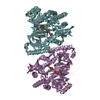
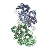
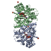
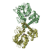
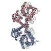
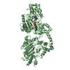
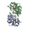
| Title | HUMAN GLUTATHIONE SYNTHETASE | ||||||
 Components Components | PROTEIN (GLUTATHIONE SYNTHETASE) | ||||||
 Keywords Keywords | AMINE/CARBOXYLATE LIGASE / AMINE-CARBOXYLATE LIGASE complex | ||||||
| Function / homology |  Function and homology information Function and homology informationDefective GSS causes GSS deficiency /  glutathione synthase / glutathione synthase activity / Glutathione synthesis and recycling / glutathione binding / amino acid metabolic process / response to cadmium ion / nervous system development / response to oxidative stress / magnesium ion binding ...Defective GSS causes GSS deficiency / glutathione synthase / glutathione synthase activity / Glutathione synthesis and recycling / glutathione binding / amino acid metabolic process / response to cadmium ion / nervous system development / response to oxidative stress / magnesium ion binding / protein homodimerization activity / extracellular exosome / ATP binding / identical protein binding / cytosol glutathione synthase / glutathione synthase activity / Glutathione synthesis and recycling / glutathione binding / amino acid metabolic process / response to cadmium ion / nervous system development / response to oxidative stress / magnesium ion binding ...Defective GSS causes GSS deficiency / glutathione synthase / glutathione synthase activity / Glutathione synthesis and recycling / glutathione binding / amino acid metabolic process / response to cadmium ion / nervous system development / response to oxidative stress / magnesium ion binding / protein homodimerization activity / extracellular exosome / ATP binding / identical protein binding / cytosolSimilarity search - Function | ||||||
| Biological species |  Homo sapiens (human) Homo sapiens (human) | ||||||
| Method | X-RAY DIFFRACTION / SYNCHROTRON / MIR / Resolution: 2.1 Å | ||||||
 Authors Authors | Polekhina, G. / Board, P. / Rossjohn, J. / Parker, M.W. | ||||||
 Citation Citation | Journal: EMBO J. / Year: 1999 Title: Molecular basis of glutathione synthetase deficiency and a rare gene permutation event. Authors: Polekhina, G. / Board, P.G. / Gali, R.R. / Rossjohn, J. / Parker, M.W. #1: Journal: Biochem.J. / Year: 1995Title: Sequencing and Expression of a Cdna for Human Glutathione Synthetase Authors: Gali, R.R. / Board, P.G. | ||||||
| History |
|
- Structure visualization
| Structure viewer | Molecule: MolmilJmol/JSmol |
|---|
- Downloads & links
-Download
| PDBx/mmCIF format | 2hgs.cif.gz | 112.1 KB | Display | PDBx/mmCIF format |
|---|---|---|---|---|
| PDB format | pdb2hgs.ent.gz | 85 KB | Display | PDB format |
| PDBx/mmJSON format | 2hgs.json.gz | Tree view | PDBx/mmJSON format | |
| Others |  Other downloads Other downloads |
-Validation report
| Arichive directory | https://data.pdbj.org/pub/pdb/validation_reports/hg/2hgsftp://data.pdbj.org/pub/pdb/validation_reports/hg/2hgs | HTTPS FTP |
|---|
-Related structure data
| Similar structure data |
|---|


-Links
 PDBj
PDBj


- Assembly
| Deposited unit | 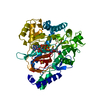
| ||||||||
|---|---|---|---|---|---|---|---|---|---|
| 1 | 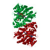
| ||||||||
| Unit cell |
|
-Components
-Protein , 1 types, 1 molecules A
| #1: Protein | Mass: 52443.621 Da / Num. of mol.: 1 Source method: isolated from a genetically manipulated source Source: (gene. exp.) Homo sapiens (human) / Description: CDNA / Organ: BRAIN / Plasmid: PRG1 / Production host:  Escherichia coli (E. coli) / Strain (production host): TG1 / References: UniProt: P48637, glutathione synthase Escherichia coli (E. coli) / Strain (production host): TG1 / References: UniProt: P48637, glutathione synthase |
|---|
-Non-polymers , 5 types, 236 molecules 


| #2: Chemical |  Mass: 24.305 Da / Num. of mol.: 2 / Source method: obtained synthetically / Formula: Mg Mass: 24.305 Da / Num. of mol.: 2 / Source method: obtained synthetically / Formula: Mg#3: Chemical | Sulfate Mass: 96.063 Da / Num. of mol.: 2 / Source method: obtained synthetically / Formula: SO4 Mass: 96.063 Da / Num. of mol.: 2 / Source method: obtained synthetically / Formula: SO4#4: Chemical | ChemComp-ADP / | Adenosine diphosphate Mass: 427.201 Da / Num. of mol.: 1 / Source method: obtained synthetically / Formula: C10H15N5O10P2 / Comment: ADP, energy-carrying molecule*YM Mass: 427.201 Da / Num. of mol.: 1 / Source method: obtained synthetically / Formula: C10H15N5O10P2 / Comment: ADP, energy-carrying molecule*YM#5: Chemical | ChemComp-GSH / | Glutathione Mass: 307.323 Da / Num. of mol.: 1 / Source method: obtained synthetically / Formula: C10H17N3O6S Mass: 307.323 Da / Num. of mol.: 1 / Source method: obtained synthetically / Formula: C10H17N3O6S#6: Water | ChemComp-HOH / | WaterMass: 18.015 Da / Num. of mol.: 230 / Source method: isolated from a natural source / Formula: H2O |
|---|
-Experimental details
-Experiment
| Experiment | Method: X-RAY DIFFRACTION / Number of used crystals: 2 |
|---|
- Sample preparation
| Crystal | Density Matthews: 3.34 Å3/Da / Density % sol: 63.2 % | ||||||||||||||||||||||||||||||||||||||||||||||||||||||||||||||||||
|---|---|---|---|---|---|---|---|---|---|---|---|---|---|---|---|---|---|---|---|---|---|---|---|---|---|---|---|---|---|---|---|---|---|---|---|---|---|---|---|---|---|---|---|---|---|---|---|---|---|---|---|---|---|---|---|---|---|---|---|---|---|---|---|---|---|---|---|
| Crystal grow | pH: 6 / Details: pH 6.0 | ||||||||||||||||||||||||||||||||||||||||||||||||||||||||||||||||||
| Crystal grow | *PLUS Temperature: 22 ℃ / pH: 7.5 / Method: vapor diffusion, hanging drop | ||||||||||||||||||||||||||||||||||||||||||||||||||||||||||||||||||
| Components of the solutions | *PLUS
|
-Data collection
| Diffraction | Mean temperature: 100 K |
|---|---|
| Diffraction source | Source: SYNCHROTRON / Site: APS  / Beamline: 14-BM-C / Wavelength: 1.037 / Beamline: 14-BM-C / Wavelength: 1.037 |
| Radiation | Protocol: SINGLE WAVELENGTH / Monochromatic (M) / Laue (L): M / Scattering type: x-ray |
| Radiation wavelength | Wavelength: 1.037 Å / Relative weight: 1 |
| Reflection | Resolution: 2.1→20 Å / Num. obs: 40495 / % possible obs: 95.9 % / Redundancy: 4 % / Rmerge(I) obs: 0.057 |
| Reflection | *PLUS Num. measured all: 201501 |
| Reflection shell | *PLUS Highest resolution: 2.1 Å / Lowest resolution: 2.2 Å / % possible obs: 95.9 % / Rmerge(I) obs: 0.197 / Mean I/σ(I) obs: 1.7 |
- Processing
| Software | Name: REFMAC / Classification: refinement | ||||||||||||||||||||||||||||||||||||||||||||||||||||||||||||||||||||||||||||||||||||
|---|---|---|---|---|---|---|---|---|---|---|---|---|---|---|---|---|---|---|---|---|---|---|---|---|---|---|---|---|---|---|---|---|---|---|---|---|---|---|---|---|---|---|---|---|---|---|---|---|---|---|---|---|---|---|---|---|---|---|---|---|---|---|---|---|---|---|---|---|---|---|---|---|---|---|---|---|---|---|---|---|---|---|---|---|---|
| Refinement | Method to determine structure: MIR / Resolution: 2.1→20 Å / Cross valid method: THROUGOUT / σ(F): 0
| ||||||||||||||||||||||||||||||||||||||||||||||||||||||||||||||||||||||||||||||||||||
| Refinement step | Cycle: LAST / Resolution: 2.1→20 Å
| ||||||||||||||||||||||||||||||||||||||||||||||||||||||||||||||||||||||||||||||||||||
| Refine LS restraints |
| ||||||||||||||||||||||||||||||||||||||||||||||||||||||||||||||||||||||||||||||||||||
| Refinement | *PLUS Rfactor obs: 0.218 | ||||||||||||||||||||||||||||||||||||||||||||||||||||||||||||||||||||||||||||||||||||
| Solvent computation | *PLUS | ||||||||||||||||||||||||||||||||||||||||||||||||||||||||||||||||||||||||||||||||||||
| Displacement parameters | *PLUS |
