 Movie
Movie Controller


+ Open data

- Basic information
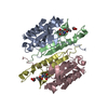
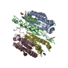
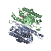
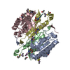
| Entry | Database: PDB / ID: 2h65 | ||||||
|---|---|---|---|---|---|---|---|
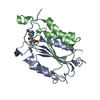
| Title | Crystal strusture of caspase-3 with inhibitor Ac-VDVAD-Cho | ||||||
 Components Components |
| ||||||
 Keywords Keywords | HYDROLASE/HYDROLASE INHIBITOR /  ENZYME CATALYSIS / CYSTEINE PROTEASE / APOPTOSIS / INDUCED FIT / HYDROLASE-HYDROLASE INHIBITOR COMPLEX ENZYME CATALYSIS / CYSTEINE PROTEASE / APOPTOSIS / INDUCED FIT / HYDROLASE-HYDROLASE INHIBITOR COMPLEX | ||||||
| Function / homology |  Function and homology informationcaspase-3 / Stimulation of the cell death response by PAK-2p34 / phospholipase A2 activator activity / anterior neural tube closure / intrinsic apoptotic signaling pathway in response to osmotic stress / leukocyte apoptotic process / positive regulation of pyroptotic inflammatory response / glial cell apoptotic process / NADE modulates death signalling / luteolysis ...caspase-3 / Stimulation of the cell death response by PAK-2p34 / phospholipase A2 activator activity / anterior neural tube closure / intrinsic apoptotic signaling pathway in response to osmotic stress / leukocyte apoptotic process / positive regulation of pyroptotic inflammatory response / glial cell apoptotic process / NADE modulates death signalling / luteolysis / response to cobalt ion / cysteine-type endopeptidase activity involved in apoptotic signaling pathway / death-inducing signaling complex / cyclin-dependent protein serine/threonine kinase inhibitor activity / cellular response to staurosporine / Apoptosis induced DNA fragmentation / Apoptotic cleavage of cell adhesion proteins / cysteine-type endopeptidase activity involved in execution phase of apoptosis / Caspase activation via Dependence Receptors in the absence of ligand / death receptor binding / SMAC, XIAP-regulated apoptotic response / axonal fasciculation / Activation of caspases through apoptosome-mediated cleavage / Signaling by Hippo / SMAC (DIABLO) binds to IAPs / SMAC(DIABLO)-mediated dissociation of IAP:caspase complexes / cysteine-type endopeptidase activity involved in apoptotic process / fibroblast apoptotic process / execution phase of apoptosis / negative regulation of cytokine production / epithelial cell apoptotic process / platelet formation / Other interleukin signaling / positive regulation of amyloid-beta formation / pyroptotic inflammatory response / Apoptotic cleavage of cellular proteins / negative regulation of B cell proliferation / T cell homeostasis / negative regulation of activated T cell proliferation / neurotrophin TRK receptor signaling pathway / B cell homeostasis / protein maturation / negative regulation of cell cycle / response to X-ray / Caspase-mediated cleavage of cytoskeletal proteins / regulation of macroautophagy / response to amino acid / cell fate commitment / Pyroptosis / response to tumor necrosis factor / response to glucose / response to UV / response to glucocorticoid / keratinocyte differentiation / striated muscle cell differentiation / Degradation of the extracellular matrix / intrinsic apoptotic signaling pathway / erythrocyte differentiation / response to nicotine / apoptotic signaling pathway / hippocampus development / sensory perception of sound / protein catabolic process / regulation of protein stability / response to hydrogen peroxide / protein processing / neuron differentiation / response to wounding / positive regulation of neuron apoptotic process / response to estradiol / heart development / peptidase activity / neuron apoptotic process / protease binding / response to lipopolysaccharide / aspartic-type endopeptidase activity / response to hypoxia / learning or memory / response to xenobiotic stimulus / positive regulation of apoptotic process / cysteine-type endopeptidase activity / neuronal cell body / apoptotic process / DNA damage response / protein-containing complex binding / proteolysis / nucleoplasm / nucleus / cytosol / cytoplasm Function and homology informationcaspase-3 / Stimulation of the cell death response by PAK-2p34 / phospholipase A2 activator activity / anterior neural tube closure / intrinsic apoptotic signaling pathway in response to osmotic stress / leukocyte apoptotic process / positive regulation of pyroptotic inflammatory response / glial cell apoptotic process / NADE modulates death signalling / luteolysis ...caspase-3 / Stimulation of the cell death response by PAK-2p34 / phospholipase A2 activator activity / anterior neural tube closure / intrinsic apoptotic signaling pathway in response to osmotic stress / leukocyte apoptotic process / positive regulation of pyroptotic inflammatory response / glial cell apoptotic process / NADE modulates death signalling / luteolysis / response to cobalt ion / cysteine-type endopeptidase activity involved in apoptotic signaling pathway / death-inducing signaling complex / cyclin-dependent protein serine/threonine kinase inhibitor activity / cellular response to staurosporine / Apoptosis induced DNA fragmentation / Apoptotic cleavage of cell adhesion proteins / cysteine-type endopeptidase activity involved in execution phase of apoptosis / Caspase activation via Dependence Receptors in the absence of ligand / death receptor binding / SMAC, XIAP-regulated apoptotic response / axonal fasciculation / Activation of caspases through apoptosome-mediated cleavage / Signaling by Hippo / SMAC (DIABLO) binds to IAPs / SMAC(DIABLO)-mediated dissociation of IAP:caspase complexes / cysteine-type endopeptidase activity involved in apoptotic process / fibroblast apoptotic process / execution phase of apoptosis / negative regulation of cytokine production / epithelial cell apoptotic process / platelet formation / Other interleukin signaling / positive regulation of amyloid-beta formation / pyroptotic inflammatory response / Apoptotic cleavage of cellular proteins / negative regulation of B cell proliferation / T cell homeostasis / negative regulation of activated T cell proliferation / neurotrophin TRK receptor signaling pathway / B cell homeostasis / protein maturation / negative regulation of cell cycle / response to X-ray / Caspase-mediated cleavage of cytoskeletal proteins / regulation of macroautophagy / response to amino acid / cell fate commitment / Pyroptosis / response to tumor necrosis factor / response to glucose / response to UV / response to glucocorticoid / keratinocyte differentiation / striated muscle cell differentiation / Degradation of the extracellular matrix / intrinsic apoptotic signaling pathway / erythrocyte differentiation / response to nicotine / apoptotic signaling pathway / hippocampus development / sensory perception of sound / protein catabolic process / regulation of protein stability / response to hydrogen peroxide / protein processing / neuron differentiation / response to wounding / positive regulation of neuron apoptotic process / response to estradiol / heart development / peptidase activity / neuron apoptotic process / protease binding / response to lipopolysaccharide / aspartic-type endopeptidase activity / response to hypoxia / learning or memory / response to xenobiotic stimulus / positive regulation of apoptotic process / cysteine-type endopeptidase activity / neuronal cell body / apoptotic process / DNA damage response / protein-containing complex binding / proteolysis / nucleoplasm / nucleus / cytosol / cytoplasmSimilarity search - Function | ||||||
| Biological species |  Homo sapiens (human) Homo sapiens (human) | ||||||
| Method | X-RAY DIFFRACTION / SYNCHROTRON / MOLECULAR REPLACEMENT / Resolution: 2.3 Å | ||||||
 Authors Authors | Fang, B. / Boross, P.I. / Tozser, J. / Weber, I.T. | ||||||
 Citation Citation | Journal: J.Mol.Biol. / Year: 2006 Title: Structural and kinetic analysis of caspase-3 reveals role for s5 binding site in substrate recognition Authors: Fang, B. / Boross, P.I. / Tozser, J. / Weber, I.T. | ||||||
| History |
|
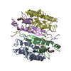
- Structure visualization
| Structure viewer | Molecule: MolmilJmol/JSmol |
|---|
- Downloads & links
-Download
| PDBx/mmCIF format | 2h65.cif.gz | 110 KB | Display | PDBx/mmCIF format |
|---|---|---|---|---|
| PDB format | pdb2h65.ent.gz | 83.6 KB | Display | PDB format |
| PDBx/mmJSON format | 2h65.json.gz | Tree view | PDBx/mmJSON format | |
| Others |  Other downloads Other downloads |
-Validation report
| Arichive directory | https://data.pdbj.org/pub/pdb/validation_reports/h6/2h65ftp://data.pdbj.org/pub/pdb/validation_reports/h6/2h65 | HTTPS FTP |
|---|
-Related structure data
| Related structure data | 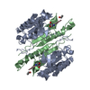 2h5iC 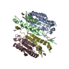 2h5jC  1cp3S C: citing same article ( S: Starting model for refinement |
|---|---|
| Similar structure data |

-Links
 PDBj
PDBj


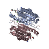
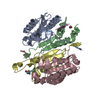
- Assembly
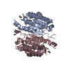
| Deposited unit | 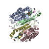
| ||||||||
|---|---|---|---|---|---|---|---|---|---|
| 1 |
| ||||||||
| Unit cell |
|
-Components
| #1: Protein | / E.C.3.4.22.- / CASP-3 / Apopain / Cysteine protease CPP32 / Yama protein / CPP-32 / SREBP cleavage activity 1 / SCA-1 Mass: 16524.814 Da / Num. of mol.: 2 / Fragment: residues 29-174 Source method: isolated from a genetically manipulated source Source: (gene. exp.) Homo sapiens (human) / Gene: CASP3, CPP32 / Plasmid: pET23b / Species (production host): Escherichia coli / Production host:  Escherichia coli BL21 (bacteria) / Strain (production host): BL21 Escherichia coli BL21 (bacteria) / Strain (production host): BL21References: UniProt: P42574, Hydrolases; Acting on peptide bonds (peptidases); Cysteine endopeptidases#2: Protein | / E.C.3.4.22.-Mass: 11257.953 Da / Num. of mol.: 2 / Fragment: residues 184-277 Source method: isolated from a genetically manipulated source Source: (gene. exp.) Homo sapiens (human) / Gene: CASP3, CPP32 / Plasmid: pET23b / Species (production host): Escherichia coli / Production host: Escherichia coli BL21 (bacteria) / Strain (production host): BL21References: UniProt: P42574, Hydrolases; Acting on peptide bonds (peptidases); Cysteine endopeptidases#3: Protein/peptide |   Type: Peptide-like / Class: Inhibitor / Mass: 529.584 Da / Num. of mol.: 2 / Source method: obtained synthetically / References: Caspase-2 Inhibitor; Ac-VDVAD-Cho Type: Peptide-like / Class: Inhibitor / Mass: 529.584 Da / Num. of mol.: 2 / Source method: obtained synthetically / References: Caspase-2 Inhibitor; Ac-VDVAD-Cho#4: Water | ChemComp-HOH / | Water Mass: 18.015 Da / Num. of mol.: 86 / Source method: isolated from a natural source / Formula: H2O Mass: 18.015 Da / Num. of mol.: 86 / Source method: isolated from a natural source / Formula: H2OCompound details | THE INHIBITOR IS COVALENTLY | |
|---|
-Experimental details
-Experiment
| Experiment | Method: X-RAY DIFFRACTION / Number of used crystals: 1 |
|---|
- Sample preparation
| Crystal | Density Matthews: 2.73 Å3/Da / Density % sol: 54.89 % |
|---|---|
| Crystal grow | Temperature: 298 K / Method: vapor diffusion, hanging drop / pH: 6.5 Details: 100 mM sodium citrate, 5% glycerol, 10 mM dithiothreitol and 14-18% PEG6000, pH 6.5, VAPOR DIFFUSION, HANGING DROP, temperature 298K |
-Data collection
| Diffraction | Mean temperature: 100 K |
|---|---|
| Diffraction source | Source: SYNCHROTRON / Site: APS  / Beamline: 22-ID / Wavelength: 1 Å / Beamline: 22-ID / Wavelength: 1 Å |
| Detector | Type: MARMOSAIC 300 mm CCD / Detector: CCD / Date: Mar 15, 2005 |
| Radiation | Monochromator: Si 220 / Protocol: SINGLE WAVELENGTH / Monochromatic (M) / Laue (L): M / Scattering type: x-ray |
| Radiation wavelength | Wavelength: 1 Å / Relative weight: 1 |
| Reflection | Resolution: 2.2→50 Å / Num. all: 34990 / Num. obs: 34990 / % possible obs: 97.6 % / Observed criterion σ(F): 0 / Observed criterion σ(I): 0 / Redundancy: 3.2 % / Biso Wilson estimate: 24.5 Å2 / Rmerge(I) obs: 0.088 / Net I/σ(I): 11 |
| Reflection shell | Resolution: 2.2→2.3 Å / Redundancy: 2.8 % / Rmerge(I) obs: 0.297 / Mean I/σ(I) obs: 4 / % possible all: 98.1 |
- Processing
| Software |
| ||||||||||||||||||||||||||||||||||||||||||||||||||||||||||||||||||||||||||||||||||||||||||||||||||||||||||||||||||||||||||||||||||||||||||||||||||||||||||||
|---|---|---|---|---|---|---|---|---|---|---|---|---|---|---|---|---|---|---|---|---|---|---|---|---|---|---|---|---|---|---|---|---|---|---|---|---|---|---|---|---|---|---|---|---|---|---|---|---|---|---|---|---|---|---|---|---|---|---|---|---|---|---|---|---|---|---|---|---|---|---|---|---|---|---|---|---|---|---|---|---|---|---|---|---|---|---|---|---|---|---|---|---|---|---|---|---|---|---|---|---|---|---|---|---|---|---|---|---|---|---|---|---|---|---|---|---|---|---|---|---|---|---|---|---|---|---|---|---|---|---|---|---|---|---|---|---|---|---|---|---|---|---|---|---|---|---|---|---|---|---|---|---|---|---|---|---|---|
| Refinement | Method to determine structure: MOLECULAR REPLACEMENT Starting model: 1CP3 Resolution: 2.3→10 Å / FOM work R set: 0.833 / Isotropic thermal model: Isotropic Cross valid method: maximum likelihood target using amplitudes σ(F): 0 / Stereochemistry target values: Engh & Huber
| ||||||||||||||||||||||||||||||||||||||||||||||||||||||||||||||||||||||||||||||||||||||||||||||||||||||||||||||||||||||||||||||||||||||||||||||||||||||||||||
| Solvent computation | Bsol: 40.134 Å2 | ||||||||||||||||||||||||||||||||||||||||||||||||||||||||||||||||||||||||||||||||||||||||||||||||||||||||||||||||||||||||||||||||||||||||||||||||||||||||||||
| Displacement parameters | Biso mean: 29.896 Å2
| ||||||||||||||||||||||||||||||||||||||||||||||||||||||||||||||||||||||||||||||||||||||||||||||||||||||||||||||||||||||||||||||||||||||||||||||||||||||||||||
| Refinement step | Cycle: LAST / Resolution: 2.3→10 Å
| ||||||||||||||||||||||||||||||||||||||||||||||||||||||||||||||||||||||||||||||||||||||||||||||||||||||||||||||||||||||||||||||||||||||||||||||||||||||||||||
| Refine LS restraints |
| ||||||||||||||||||||||||||||||||||||||||||||||||||||||||||||||||||||||||||||||||||||||||||||||||||||||||||||||||||||||||||||||||||||||||||||||||||||||||||||
| LS refinement shell | Refine-ID: X-RAY DIFFRACTION / Total num. of bins used: 25
| ||||||||||||||||||||||||||||||||||||||||||||||||||||||||||||||||||||||||||||||||||||||||||||||||||||||||||||||||||||||||||||||||||||||||||||||||||||||||||||
| Xplor file |
|
