 Movie
Movie Controller

[English] 日本語
 Yorodumi
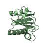
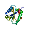
- PDB-2gte: Drosophila OBP LUSH bound to attractant pheromone 11-cis-vaccenyl... -
+ Open data

- Basic information
| Entry | Database: PDB / ID: 2gte | ||||||
|---|---|---|---|---|---|---|---|
| Title | Drosophila OBP LUSH bound to attractant pheromone 11-cis-vaccenyl acetate | ||||||
 Components Components | General odorant-binding protein lush | ||||||
 Keywords Keywords |  TRANSPORT PROTEIN / pheromone binding protein / odorant binding protein / pheromone TRANSPORT PROTEIN / pheromone binding protein / odorant binding protein / pheromone | ||||||
| Function / homology |  Function and homology information Function and homology informationdiphenyl phthalate binding / courtship behavior / dibutyl phthalate binding / response to pheromone / pheromone binding / olfactory behavior / odorant binding / sensory perception of smell / response to ethanol / extracellular regionSimilarity search - Function | ||||||
| Biological species |  Drosophila melanogaster (fruit fly) Drosophila melanogaster (fruit fly) | ||||||
| Method | X-RAY DIFFRACTION / SYNCHROTRON / MOLECULAR REPLACEMENT / Resolution: 1.4 Å | ||||||
 Authors Authors | Laughlin, J.D. / Ha, T. / Smith, D.P. / Jones, D.N.M. | ||||||
 Citation Citation | Journal: Cell(Cambridge,Mass.) / Year: 2008 Title: Activation of pheromone-sensitive neurons is mediated by conformational activation of pheromone-binding protein Authors: Laughlin, J.D. / Ha, T.S. / Jones, D.N. / Smith, D.P. | ||||||
| History |
|
- Structure visualization
| Structure viewer | Molecule: MolmilJmol/JSmol |
|---|
- Downloads & links
-Download
| PDBx/mmCIF format | 2gte.cif.gz | 75.2 KB | Display | PDBx/mmCIF format |
|---|---|---|---|---|
| PDB format | pdb2gte.ent.gz | 56.2 KB | Display | PDB format |
| PDBx/mmJSON format | 2gte.json.gz | Tree view | PDBx/mmJSON format | |
| Others |  Other downloads Other downloads |
-Validation report
| Arichive directory | https://data.pdbj.org/pub/pdb/validation_reports/gt/2gteftp://data.pdbj.org/pub/pdb/validation_reports/gt/2gte | HTTPS FTP |
|---|
-Related structure data


-Links
 PDBj
PDBj- Assembly
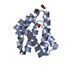
| Deposited unit | 
| ||||||||
|---|---|---|---|---|---|---|---|---|---|
| 1 | 
| ||||||||
| 2 | 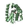
| ||||||||
| Unit cell |
|
-Components
| #1: Protein | Mass: 14215.508 Da / Num. of mol.: 2 / Fragment: Residues 30-153 Source method: isolated from a genetically manipulated source Source: (gene. exp.) Drosophila melanogaster (fruit fly) / Gene: lush / Plasmid: pET13 / Production host:  Escherichia coli (E. coli) / Strain (production host): BL21 (DE3) / References: UniProt: O02372 Escherichia coli (E. coli) / Strain (production host): BL21 (DE3) / References: UniProt: O02372#2: Chemical | ChemComp-PO4 / | Phosphate  Mass: 94.971 Da / Num. of mol.: 1 / Source method: obtained synthetically / Formula: PO4 Mass: 94.971 Da / Num. of mol.: 1 / Source method: obtained synthetically / Formula: PO4#3: Chemical | Vaccenyl acetate  Mass: 310.515 Da / Num. of mol.: 2 / Source method: obtained synthetically / Formula: C20H38O2 Mass: 310.515 Da / Num. of mol.: 2 / Source method: obtained synthetically / Formula: C20H38O2#4: Water | ChemComp-HOH / | Water Mass: 18.015 Da / Num. of mol.: 416 / Source method: isolated from a natural source / Formula: H2O Mass: 18.015 Da / Num. of mol.: 416 / Source method: isolated from a natural source / Formula: H2O |
|---|
-Experimental details
-Experiment
| Experiment | Method: X-RAY DIFFRACTION / Number of used crystals: 1 |
|---|
- Sample preparation
| Crystal | Density Matthews: 2.46 Å3/Da / Density % sol: 49.93 % |
|---|---|
| Crystal grow | Temperature: 277 K / Method: vapor diffusion, hanging drop / pH: 8.5 Details: 1:1 protein/well sol. (4ul total) over 0.5 mL well, pH 8.5, VAPOR DIFFUSION, HANGING DROP, temperature 277K |
-Data collection
| Diffraction | Mean temperature: 95 K |
|---|---|
| Diffraction source | Source: SYNCHROTRON / Site: ALS  / Beamline: 4.2.2 / Wavelength: 1.127 Å / Beamline: 4.2.2 / Wavelength: 1.127 Å |
| Detector | Detector: CCD |
| Radiation | Protocol: SINGLE WAVELENGTH / Monochromatic (M) / Laue (L): M / Scattering type: x-ray |
| Radiation wavelength | Wavelength: 1.127 Å / Relative weight: 1 |
| Reflection | Resolution: 1.4→41.2 Å / Num. obs: 55876 / % possible obs: 99.9 % / Redundancy: 6.68 % / Rmerge(I) obs: 0.078 / Net I/σ(I): 14 |
| Reflection shell | Resolution: 1.4→1.45 Å / Redundancy: 5.33 % / Rmerge(I) obs: 0.311 / Mean I/σ(I) obs: 4.2 / % possible all: 99.9 |
- Processing
| Software |
| ||||||||||||||||||||||||||||||||||||||||||||||||||||||||||||||||||||||||||||||||||||||||||||||||||||||||||||||||||||||||||||||||||||||||||||||||||||||||||||||||||||||||||
|---|---|---|---|---|---|---|---|---|---|---|---|---|---|---|---|---|---|---|---|---|---|---|---|---|---|---|---|---|---|---|---|---|---|---|---|---|---|---|---|---|---|---|---|---|---|---|---|---|---|---|---|---|---|---|---|---|---|---|---|---|---|---|---|---|---|---|---|---|---|---|---|---|---|---|---|---|---|---|---|---|---|---|---|---|---|---|---|---|---|---|---|---|---|---|---|---|---|---|---|---|---|---|---|---|---|---|---|---|---|---|---|---|---|---|---|---|---|---|---|---|---|---|---|---|---|---|---|---|---|---|---|---|---|---|---|---|---|---|---|---|---|---|---|---|---|---|---|---|---|---|---|---|---|---|---|---|---|---|---|---|---|---|---|---|---|---|---|---|---|---|---|
| Refinement | Method to determine structure: MOLECULAR REPLACEMENT Starting model: Apo-LUSH Resolution: 1.4→41.2 Å / Cor.coef. Fo:Fc: 0.96 / Cor.coef. Fo:Fc free: 0.946 / Cross valid method: THROUGHOUT / ESU R: 0.065 / ESU R Free: 0.067 / Stereochemistry target values: MAXIMUM LIKELIHOOD
| ||||||||||||||||||||||||||||||||||||||||||||||||||||||||||||||||||||||||||||||||||||||||||||||||||||||||||||||||||||||||||||||||||||||||||||||||||||||||||||||||||||||||||
| Solvent computation | Ion probe radii: 0.8 Å / Shrinkage radii: 0.8 Å / VDW probe radii: 1.2 Å / Solvent model: BABINET MODEL WITH MASK | ||||||||||||||||||||||||||||||||||||||||||||||||||||||||||||||||||||||||||||||||||||||||||||||||||||||||||||||||||||||||||||||||||||||||||||||||||||||||||||||||||||||||||
| Displacement parameters | Biso mean: 12.969 Å2
| ||||||||||||||||||||||||||||||||||||||||||||||||||||||||||||||||||||||||||||||||||||||||||||||||||||||||||||||||||||||||||||||||||||||||||||||||||||||||||||||||||||||||||
| Refinement step | Cycle: LAST / Resolution: 1.4→41.2 Å
| ||||||||||||||||||||||||||||||||||||||||||||||||||||||||||||||||||||||||||||||||||||||||||||||||||||||||||||||||||||||||||||||||||||||||||||||||||||||||||||||||||||||||||
| Refine LS restraints |
| ||||||||||||||||||||||||||||||||||||||||||||||||||||||||||||||||||||||||||||||||||||||||||||||||||||||||||||||||||||||||||||||||||||||||||||||||||||||||||||||||||||||||||
| LS refinement shell | Resolution: 1.4→1.436 Å / Total num. of bins used: 20
|
