 Movie
Movie Controller

[English] 日本語
 Yorodumi
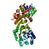
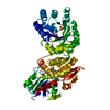
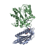
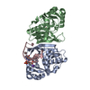
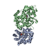
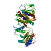
- PDB-2gpt: Crystal structure of Arabidopsis Dehydroquinate dehydratase-shiki... -
+ Open data

- Basic information
| Entry | Database: PDB / ID: 2gpt | ||||||
|---|---|---|---|---|---|---|---|
| Title | Crystal structure of Arabidopsis Dehydroquinate dehydratase-shikimate dehydrogenase in complex with tartrate and shikimate | ||||||
 Components Components | 3-dehydroquinate dehydratase/ shikimate 5-dehydrogenase | ||||||
 Keywords Keywords |  OXIDOREDUCTASE / LYASE / Type I dehydroquinate dehydratase / AroE / shikimate dehydrogenase / Arabidopsis thaliana / shikimate / tartrate / bifunctional enzyme OXIDOREDUCTASE / LYASE / Type I dehydroquinate dehydratase / AroE / shikimate dehydrogenase / Arabidopsis thaliana / shikimate / tartrate / bifunctional enzyme | ||||||
| Function / homology |  Function and homology information Function and homology informationshikimate dehydrogenase (NADP+) / shikimate metabolic process / shikimate 3-dehydrogenase (NADP+) activity / embryo development ending in seed dormancy / 3-dehydroquinate dehydratase / 3-dehydroquinate dehydratase activity / chorismate biosynthetic process / chloroplast stroma / aromatic amino acid family biosynthetic process / amino acid biosynthetic process ...shikimate dehydrogenase (NADP+) / shikimate metabolic process / shikimate 3-dehydrogenase (NADP+) activity / embryo development ending in seed dormancy / 3-dehydroquinate dehydratase / 3-dehydroquinate dehydratase activity / chorismate biosynthetic process / chloroplast stroma / aromatic amino acid family biosynthetic process / amino acid biosynthetic process / chloroplast / NADP bindingSimilarity search - Function | ||||||
| Biological species |  Arabidopsis thaliana (thale cress) Arabidopsis thaliana (thale cress) | ||||||
| Method | X-RAY DIFFRACTION / SYNCHROTRON / SAD / Resolution: 1.95 Å | ||||||
 Authors Authors | Singh, S.A. / Christendat, D. | ||||||
 Citation Citation | Journal: Biochemistry / Year: 2006 Title: Structure of Arabidopsis dehydroquinate dehydratase-shikimate dehydrogenase and implications for metabolic channeling in the shikimate pathway Authors: Singh, S.A. / Christendat, D. | ||||||
| History |
|
- Structure visualization
| Structure viewer | Molecule: MolmilJmol/JSmol |
|---|
- Downloads & links
-Download
| PDBx/mmCIF format | 2gpt.cif.gz | 115.5 KB | Display | PDBx/mmCIF format |
|---|---|---|---|---|
| PDB format | pdb2gpt.ent.gz | 87.6 KB | Display | PDB format |
| PDBx/mmJSON format | 2gpt.json.gz | Tree view | PDBx/mmJSON format | |
| Others |  Other downloads Other downloads |
-Validation report
| Arichive directory | https://data.pdbj.org/pub/pdb/validation_reports/gp/2gptftp://data.pdbj.org/pub/pdb/validation_reports/gp/2gpt | HTTPS FTP |
|---|
-Related structure data
| Similar structure data |
|---|


-Links
 PDBj
PDBj

- Assembly
| Deposited unit | 
| ||||||||
|---|---|---|---|---|---|---|---|---|---|
| 1 |
| ||||||||
| Unit cell |
| ||||||||
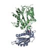
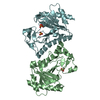
| Details | The biological assembly is a monomer |
-Components
| #1: Protein | Mass: 57088.957 Da / Num. of mol.: 1 Source method: isolated from a genetically manipulated source Source: (gene. exp.) Arabidopsis thaliana (thale cress) / Production host:  Escherichia coli (E. coli) Escherichia coli (E. coli)References: UniProt: Q9SQT8, 3-dehydroquinate dehydratase, shikimate dehydrogenase (NADP+) | ||||||
|---|---|---|---|---|---|---|---|
| #2: Chemical | Sulfate  Mass: 96.063 Da / Num. of mol.: 2 / Source method: obtained synthetically / Formula: SO4 Mass: 96.063 Da / Num. of mol.: 2 / Source method: obtained synthetically / Formula: SO4#3: Chemical | ChemComp-TLA / | Tartaric acid  Mass: 150.087 Da / Num. of mol.: 1 / Source method: obtained synthetically / Formula: C4H6O6 Mass: 150.087 Da / Num. of mol.: 1 / Source method: obtained synthetically / Formula: C4H6O6#4: Chemical | ChemComp-SKM / ( | Shikimic acid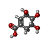  Mass: 174.151 Da / Num. of mol.: 1 / Source method: obtained synthetically / Formula: C7H10O5 Mass: 174.151 Da / Num. of mol.: 1 / Source method: obtained synthetically / Formula: C7H10O5#5: Water | ChemComp-HOH / | Water Mass: 18.015 Da / Num. of mol.: 271 / Source method: isolated from a natural source / Formula: H2O Mass: 18.015 Da / Num. of mol.: 271 / Source method: isolated from a natural source / Formula: H2O |
-Experimental details
-Experiment
| Experiment | Method: X-RAY DIFFRACTION |
|---|
- Sample preparation
| Crystal | Density Matthews: 2.76 Å3/Da / Density % sol: 55.41 % |
|---|---|
| Crystal grow | Temperature: 298 K / Method: vapor diffusion, hanging drop / pH: 5.6 Details: 0.4 mM Sodium Tartrate tetrahydrate, 0.1 M tri-Sodium Citrate dihydrate pH5.6, 2.8 M Ammonium Sulfate, VAPOR DIFFUSION, HANGING DROP, temperature 298K |
-Data collection
| Diffraction |
| |||||||||||||||
|---|---|---|---|---|---|---|---|---|---|---|---|---|---|---|---|---|
| Diffraction source |
| |||||||||||||||
| Detector | Type: ADSC QUANTUM 4 / Detector: CCD / Date: Nov 19, 2004 | |||||||||||||||
| Radiation | Protocol: SINGLE WAVELENGTH / Monochromatic (M) / Laue (L): M / Scattering type: x-ray | |||||||||||||||
| Radiation wavelength |
| |||||||||||||||
| Reflection | Resolution: 1.95→50 Å / Num. obs: 46428 / % possible obs: 99.7 % / Observed criterion σ(F): 0 / Observed criterion σ(I): 0 / Redundancy: 7.1 % / Biso Wilson estimate: 21.4 Å2 / Rmerge(I) obs: 0.058 / Rsym value: 0.044 / Net I/σ(I): 42.9 | |||||||||||||||
| Reflection shell | Highest resolution: 1.95 Å / Redundancy: 6.1 % / Rmerge(I) obs: 0.635 / Mean I/σ(I) obs: 3.3 / Rsym value: 0.537 / % possible all: 98.5 |

- Processing
| Software |
| ||||||||||||||||||||||||||||||||||||
|---|---|---|---|---|---|---|---|---|---|---|---|---|---|---|---|---|---|---|---|---|---|---|---|---|---|---|---|---|---|---|---|---|---|---|---|---|---|
| Refinement | Method to determine structure: SAD / Resolution: 1.95→28.46 Å / Rfactor Rfree error: 0.007 / Data cutoff high absF: 279246.46 / Data cutoff low absF: 0 / Isotropic thermal model: RESTRAINED / Cross valid method: THROUGHOUT / σ(F): 0 / Stereochemistry target values: Engh & Huber
| ||||||||||||||||||||||||||||||||||||
| Solvent computation | Solvent model: FLAT MODEL / Bsol: 56.1233 Å2 / ksol: 0.362598 e/Å3 | ||||||||||||||||||||||||||||||||||||
| Displacement parameters | Biso mean: 38.7 Å2
| ||||||||||||||||||||||||||||||||||||
| Refine analyze |
| ||||||||||||||||||||||||||||||||||||
| Refinement step | Cycle: LAST / Resolution: 1.95→28.46 Å
| ||||||||||||||||||||||||||||||||||||
| Refine LS restraints |
| ||||||||||||||||||||||||||||||||||||
| LS refinement shell | Resolution: 1.95→2.07 Å / Rfactor Rfree error: 0.025 / Total num. of bins used: 6
| ||||||||||||||||||||||||||||||||||||
| Xplor file |
|
