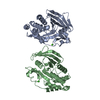
 Movie
Movie Controller


+ Open data

- Basic information
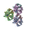
| Entry | Database: PDB / ID: 2glt | |||||||||
|---|---|---|---|---|---|---|---|---|---|---|
| Title | STRUCTURE OF ESCHERICHIA COLI GLUTATHIONE SYNTHETASE AT PH 6.0. | |||||||||
 Components Components | GLUTATHIONE BIOSYNTHETIC LIGASE | |||||||||
 Keywords Keywords |  BIOSYNTHESIS / LIGASE / GLUTATHIONE BIOSYNTHESIS LIGASE BIOSYNTHESIS / LIGASE / GLUTATHIONE BIOSYNTHESIS LIGASE | |||||||||
| Function / homology |  Function and homology informationglutathione synthase / glutathione synthase activity / glutathione biosynthetic process / protein homotetramerization / magnesium ion binding / ATP binding / identical protein binding / cytosol / cytoplasm Function and homology informationglutathione synthase / glutathione synthase activity / glutathione biosynthetic process / protein homotetramerization / magnesium ion binding / ATP binding / identical protein binding / cytosol / cytoplasmSimilarity search - Function | |||||||||
| Biological species |  Escherichia coli (E. coli) Escherichia coli (E. coli) | |||||||||
| Method | X-RAY DIFFRACTION / SYNCHROTRON / Resolution: 2.2 Å | |||||||||
 Authors Authors | Matsuda, K. / Yamaguchi, H. / Kato, H. / Nishioka, T. / Katsube, Y. / Oda, J. | |||||||||
 Citation Citation | Journal: Protein Eng. / Year: 1996 Title: Crystal structure of glutathione synthetase at optimal pH: domain architecture and structural similarity with other proteins. Authors: Matsuda, K. / Mizuguchi, K. / Nishioka, T. / Kato, H. / Go, N. / Oda, J. #1: Journal: Biochemistry / Year: 1994Title: Flexible Loop that is Novel Catalytic Machinery in a Ligase. Atomic Structure and Function of the Loopless Glutathione Synthetase Authors: Kato, H. / Tanaka, T. / Yamaguchi, H. / Hara, T. / Nishioka, T. / Katsube, Y. / Oda, J. #2: Journal: J.Am.Chem.Soc. / Year: 1994Title: Mechanism-Based Inactivation of Glutathione Synthetase by Phosphinic Acid Transition-State Analogue Authors: Hiratake, J. / Kato, H. / Oda, J. #3: Journal: Biochemistry / Year: 1993Title: Use of Adenosine (5')Polyphospho(5')Pyridoxals to Study the Substrate-Binding Region of Glutathione Synthetase from Escherichia Coli B Authors: Hibi, T. / Kato, H. / Nishioka, T. / Oda, J. / Yamaguchi, H. / Katsube, Y. / Tanizawa, K. / Fukui, T. #4: Journal: Biochemistry / Year: 1993Title: Flexibility Impaired by Mutations Revealed the Multi-Functional Roles of the Loop in Glutathione Synthetase Authors: Tanaka, T. / Yamaguchi, H. / Kato, H. / Nishioka, T. / Katsube, Y. / Oda, J. #5: Journal: J.Mol.Biol. / Year: 1993Title: Three-Dimensional Structure of the Glutathione Synthetase from Escherichia Coli B at 2.0 Angstroms Resolution Authors: Yamaguchi, H. / Kato, H. / Hata, Y. / Nishioka, T. / Kimura, A. / Oda, J. / Katsube, Y. #6: Journal: Bull.Inst.Chem.Res.,Kyoto Univ. / Year: 1993Title: Construction, Expression, and Characterization of Glutathione Synthetase Chimeras: Substitution of a Homologous Loop Peptide Region of Dihydrofolate Reductase Authors: Tanaka, T. / Sakai, T. / Chihara-Siomi, M. / Takeshima, K. / Kato, H. / Misawa, T. / Nishioka, T. / Oda, J. #7: Journal: Biochemistry / Year: 1992Title: Mutational and Proteolytic Studies on a Flexible Loop in Glutathione Synthetase from Escherichia Coli B: The Loop and Arginine 233 are Critical for the Catalytic Reaction Authors: Tanaka, T. / Kato, H. / Nishioka, T. / Oda, J. #8: Journal: Photon Factory Activity Report / Year: 1992Title: Structural Studies on Glutathione Synthetase from Escherichia Coli B Authors: Yamaguchi, H. / Kato, H. / Hata, Y. / Nishioka, T. / Oda, J. / Katsube, Y. #9: Journal: J.Mol.Biol. / Year: 1989Title: Crystallization and Preliminary X-Ray Studies of Glutathione Synthetase from Escherichia Coli B Authors: Kato, H. / Yamaguchi, H. / Hata, Y. / Nishioka, T. / Katsube, Y. / Oda, J. #10: Journal: Agric.Biol.Chem. / Year: 1989Title: Overexpression of Glutathione Synthase in Escherichia Coli Authors: Kato, H. / Kobayashi, M. / Murata, K. / Nishioka, T. / Oda, J. #11: Journal: J.Biol.Chem. / Year: 1988Title: Role of Cysteine Residues in Glutathione Synthetase from Escherchia Coli B Authors: Kato, H. / Tanaka, T. / Nishioka, T. / Kimura, A. / Oda, J. #12: Journal: Nucleic Acids Res. / Year: 1984Title: Complete Nucleotide Sequence of E.Coli Glutathione Synthetase Gsh-II Authors: Gushima, H. / Yasuda, S. / Soeda, E. / Yokota, M. / Kondo, M. / Kimura, A. | |||||||||
| History |
|
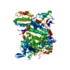
- Structure visualization
| Structure viewer | Molecule: MolmilJmol/JSmol |
|---|
- Downloads & links
-Download
| PDBx/mmCIF format | 2glt.cif.gz | 73.9 KB | Display | PDBx/mmCIF format |
|---|---|---|---|---|
| PDB format | pdb2glt.ent.gz | 55.5 KB | Display | PDB format |
| PDBx/mmJSON format | 2glt.json.gz | Tree view | PDBx/mmJSON format | |
| Others |  Other downloads Other downloads |
-Validation report
| Arichive directory | https://data.pdbj.org/pub/pdb/validation_reports/gl/2gltftp://data.pdbj.org/pub/pdb/validation_reports/gl/2glt | HTTPS FTP |
|---|
-Related structure data


-Links
 PDBj
PDBj


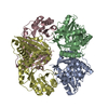
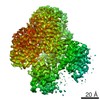
- Assembly
| Deposited unit | 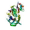
| ||||||||
|---|---|---|---|---|---|---|---|---|---|
| 1 | 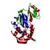
| ||||||||
| Unit cell |
| ||||||||
| Atom site foot note | 1: CIS PROLINE - PRO 90 2: VAL 113 - ASN 114 OMEGA = 357.86 PEPTIDE BOND DEVIATES SIGNIFICANTLY FROM TRANS CONFORMATION | ||||||||
| Details | SYMMETRY THE CRYSTALLOGRAPHIC SYMMETRY TRANSFORMATIONS PRESENTED BELOW GENERATE THE SUBUNITS OF THE POLYMERIC MOLECULE. APPLIED TO RESIDUES: 1 .. 316 SYMMETRY1 1 0.000000 1.000000 0.000000 0.00000 SYMMETRY2 1 1.000000 0.000000 0.000000 0.00000 SYMMETRY3 1 0.000000 0.000000 -1.000000 0.66667 SYMMETRY1 2 -1.000000 0.000000 0.000000 1.00000 SYMMETRY2 2 0.000000 -1.000000 0.000000 1.00000 SYMMETRY3 2 0.000000 0.000000 1.000000 0.00000 SYMMETRY1 3 0.000000 -1.000000 0.000000 1.00000 SYMMETRY2 3 -1.000000 0.000000 0.000000 1.00000 SYMMETRY3 3 0.000000 0.000000 -1.000000 0.66667 |
-Components
| #1: Protein | Mass: 35601.824 Da / Num. of mol.: 1 Source method: isolated from a genetically manipulated source Source: (gene. exp.) Escherichia coli (E. coli) / Strain: B / Gene: GSHII / Plasmid: PKGS00 A DERIVATIVE OF / Gene (production host): GSHII / References: UniProt: P04425, glutathione synthase |
|---|---|
| #2: Water | ChemComp-HOH / Water Mass: 18.015 Da / Num. of mol.: 92 / Source method: isolated from a natural source / Formula: H2O Mass: 18.015 Da / Num. of mol.: 92 / Source method: isolated from a natural source / Formula: H2O |
-Experimental details
-Experiment
| Experiment | Method: X-RAY DIFFRACTION / Number of used crystals: 1 |
|---|
- Sample preparation
| Crystal | Density Matthews: 2.58 Å3/Da / Density % sol: 52.26 % Description: INTENSITY DATA WERE COLLECTED WITH A WEISSENBERG CAMERA EQUIPPED WITH A CYLINDRICAL CASSETTE HAVING A 430 MM RADIUS AND THE FUJI FILM IMAGING PLATES | ||||||||||||||||||||||||
|---|---|---|---|---|---|---|---|---|---|---|---|---|---|---|---|---|---|---|---|---|---|---|---|---|---|
| Crystal grow | *PLUS Method: unknown | ||||||||||||||||||||||||
| Components of the solutions | *PLUS
|
-Data collection
| Diffraction source | Source: SYNCHROTRON / Site: Photon Factory  / Beamline: BL-6A / Wavelength: 1.04 Å / Beamline: BL-6A / Wavelength: 1.04 Å |
|---|---|
| Detector | Type: WEISSENBERG / Detector: DIFFRACTOMETER / Date: Aug 3, 1990 |
| Radiation | Monochromatic (M) / Laue (L): M / Scattering type: x-ray |
| Radiation wavelength | Wavelength: 1.04 Å / Relative weight: 1 |
| Reflection | Resolution: 2.2→77.8 Å / Num. obs: 24571 / % possible obs: 84.1 % / Observed criterion σ(I): 1 |
| Reflection | *PLUS % possible obs: 77.8 % / Rmerge(I) obs: 0.053 |
- Processing
| Software |
| ||||||||||||||||||||||||||||||||||||||||||||||||||||||||||||
|---|---|---|---|---|---|---|---|---|---|---|---|---|---|---|---|---|---|---|---|---|---|---|---|---|---|---|---|---|---|---|---|---|---|---|---|---|---|---|---|---|---|---|---|---|---|---|---|---|---|---|---|---|---|---|---|---|---|---|---|---|---|
| Refinement | Resolution: 2.2→10 Å / σ(F): 2
| ||||||||||||||||||||||||||||||||||||||||||||||||||||||||||||
| Displacement parameters | Biso mean: 21.49 Å2 | ||||||||||||||||||||||||||||||||||||||||||||||||||||||||||||
| Refinement step | Cycle: LAST / Resolution: 2.2→10 Å
| ||||||||||||||||||||||||||||||||||||||||||||||||||||||||||||
| Refine LS restraints |
| ||||||||||||||||||||||||||||||||||||||||||||||||||||||||||||
| Software | *PLUS Name: X-PLOR / Version: 3.1 / Classification: refinement | ||||||||||||||||||||||||||||||||||||||||||||||||||||||||||||
| Refinement | *PLUS Rfactor Rfree: 0.26 | ||||||||||||||||||||||||||||||||||||||||||||||||||||||||||||
| Solvent computation | *PLUS | ||||||||||||||||||||||||||||||||||||||||||||||||||||||||||||
| Displacement parameters | *PLUS | ||||||||||||||||||||||||||||||||||||||||||||||||||||||||||||
| Refine LS restraints | *PLUS
|
