 Movie
Movie Controller


+ Open data

- Basic information
| Entry | Database: PDB / ID: 2gje | ||||||
|---|---|---|---|---|---|---|---|
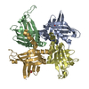
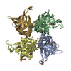
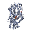
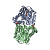
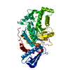
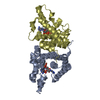
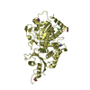
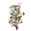
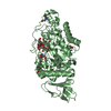
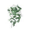
| Title | Structure of a guideRNA-binding protein complex bound to a gRNA | ||||||
 Components Components |
| ||||||
 Keywords Keywords | TRANSLATION/RNA /  guide RNA / kRNA editing / RNA binding protein / TRANSLATION-RNA COMPLEX guide RNA / kRNA editing / RNA binding protein / TRANSLATION-RNA COMPLEX | ||||||
| Function / homology |  Function and homology informationRNA modification / mRNA modification / kinetoplast / nuclear lumen / post-transcriptional regulation of gene expression / mRNA binding / regulation of DNA-templated transcription / mitochondrion / DNA binding / RNA binding Function and homology informationRNA modification / mRNA modification / kinetoplast / nuclear lumen / post-transcriptional regulation of gene expression / mRNA binding / regulation of DNA-templated transcription / mitochondrion / DNA binding / RNA bindingSimilarity search - Function | ||||||
| Biological species |  Trypanosoma brucei (eukaryote) Trypanosoma brucei (eukaryote) | ||||||
| Method | X-RAY DIFFRACTION / SYNCHROTRON / MOLECULAR REPLACEMENT / Resolution: 3.37 Å | ||||||
 Authors Authors | Schumacher, M.A. / Karamooz, E. / Zikova, A. / Trantirek, L. / Lukes, J. | ||||||
 Citation Citation | Journal: Cell(Cambridge,Mass.) / Year: 2006 Title: Crystal Structures of T. brucei MRP1/MRP2 Guide-RNA Binding Complex Reveal RNA Matchmaking Mechanism. Authors: Schumacher, M.A. / Karamooz, E. / Zikova, A. / Trantirek, L. / Lukes, J. | ||||||
| History |
|
- Structure visualization
| Structure viewer | Molecule: MolmilJmol/JSmol |
|---|
- Downloads & links
-Download
| PDBx/mmCIF format | 2gje.cif.gz | 81.2 KB | Display | PDBx/mmCIF format |
|---|---|---|---|---|
| PDB format | pdb2gje.ent.gz | 64.4 KB | Display | PDB format |
| PDBx/mmJSON format | 2gje.json.gz | Tree view | PDBx/mmJSON format | |
| Others |  Other downloads Other downloads |
-Validation report
| Arichive directory | https://data.pdbj.org/pub/pdb/validation_reports/gj/2gjeftp://data.pdbj.org/pub/pdb/validation_reports/gj/2gje | HTTPS FTP |
|---|
-Related structure data


-Links
 PDBj
PDBj


- Assembly
| Deposited unit | 
| ||||||||
|---|---|---|---|---|---|---|---|---|---|
| 1 |
| ||||||||
| Unit cell |
| ||||||||
| Details | MRP1/MRP2 form a heterotetramer with pseudo C4 symmetry ++ RNA single strand |
-Components
| #1: RNA chain | Mass: 12534.474 Da / Num. of mol.: 1 / Source method: obtained synthetically |
|---|---|
| #2: RNA chain | Mass: 1586.992 Da / Num. of mol.: 1 / Source method: obtained synthetically |
| #3: Protein | Mass: 22055.076 Da / Num. of mol.: 1 Source method: isolated from a genetically manipulated source Source: (gene. exp.) Trypanosoma brucei (eukaryote) / Strain: TREU927 / Gene: mrp1 / Plasmid: petduet-1 / Species (production host): Escherichia coli / Production host:  Escherichia coli BL21(DE3) (bacteria) / Strain (production host): BL21(DE3) / References: UniProt: Q952G2 Escherichia coli BL21(DE3) (bacteria) / Strain (production host): BL21(DE3) / References: UniProt: Q952G2 |
| #4: Protein | Mass: 21333.455 Da / Num. of mol.: 1 Source method: isolated from a genetically manipulated source Source: (gene. exp.) Trypanosoma brucei (eukaryote) / Strain: TREU927 / Gene: mrp2 / Plasmid: petduet-1 / Species (production host): Escherichia coli / Production host: Escherichia coli BL21(DE3) (bacteria) / Strain (production host): BL21(DE3) / References: UniProt: P90629 |
| #5: Water | ChemComp-HOH / Water Mass: 18.015 Da / Num. of mol.: 1 / Source method: isolated from a natural source / Formula: H2O Mass: 18.015 Da / Num. of mol.: 1 / Source method: isolated from a natural source / Formula: H2O |
-Experimental details
-Experiment
| Experiment | Method: X-RAY DIFFRACTION / Number of used crystals: 1 |
|---|
- Sample preparation
| Crystal | Density Matthews: 2.53 Å3/Da / Density % sol: 51.45 % | ||||||||||||||||||||||||||||
|---|---|---|---|---|---|---|---|---|---|---|---|---|---|---|---|---|---|---|---|---|---|---|---|---|---|---|---|---|---|
| Crystal grow | Temperature: 280 K / Method: vapor diffusion, hanging drop / pH: 7.5 Details: peg 4000, hepes, isopropanol, pH 7.5, VAPOR DIFFUSION, HANGING DROP, temperature 280K | ||||||||||||||||||||||||||||
| Components of the solutions |
|
-Data collection
| Diffraction | Mean temperature: 100 K |
|---|---|
| Diffraction source | Source: SYNCHROTRON / Site: ALS  / Beamline: 8.3.1 / Wavelength: 0.989 Å / Beamline: 8.3.1 / Wavelength: 0.989 Å |
| Detector | Type: ADSC QUANTUM 4 / Detector: CCD / Date: Dec 20, 2005 / Details: mirrors |
| Radiation | Protocol: SINGLE WAVELENGTH / Monochromatic (M) / Laue (L): M / Scattering type: x-ray |
| Radiation wavelength | Wavelength: 0.989 Å / Relative weight: 1 |
| Reflection | Resolution: 3.37→81.6 Å / Num. all: 8830 / Num. obs: 8126 / % possible obs: 92 % / Observed criterion σ(F): 0 / Observed criterion σ(I): 0 |
| Reflection shell | Resolution: 3.37→3.45 Å / % possible all: 70 |
- Processing
| Software |
| |||||||||||||||||||||||||
|---|---|---|---|---|---|---|---|---|---|---|---|---|---|---|---|---|---|---|---|---|---|---|---|---|---|---|
| Refinement | Method to determine structure: MOLECULAR REPLACEMENT / Resolution: 3.37→69.85 Å / Rfactor Rfree error: 0.012 / Data cutoff high absF: 6089525.04 / Data cutoff low absF: 0 / Isotropic thermal model: RESTRAINED / Cross valid method: THROUGHOUT / σ(F): 0 / Stereochemistry target values: Engh & Huber
| |||||||||||||||||||||||||
| Solvent computation | Solvent model: FLAT MODEL / Bsol: 53.7629 Å2 / ksol: 0.274147 e/Å3 | |||||||||||||||||||||||||
| Displacement parameters | Biso mean: 82.1 Å2
| |||||||||||||||||||||||||
| Refine analyze |
| |||||||||||||||||||||||||
| Refinement step | Cycle: LAST / Resolution: 3.37→69.85 Å
| |||||||||||||||||||||||||
| Refine LS restraints |
| |||||||||||||||||||||||||
| LS refinement shell | Resolution: 3.37→3.58 Å / Rfactor Rfree error: 0.05 / Total num. of bins used: 6
| |||||||||||||||||||||||||
| Xplor file |
|
