 Movie
Movie Controller

[English] 日本語
 Yorodumi
- PDB-2gjd: Distinct functional domains of Ubc9 dictate cell survival and res... -
+ Open data

- Basic information
| Entry | Database: PDB / ID: 2gjd | ||||||
|---|---|---|---|---|---|---|---|
| Title | Distinct functional domains of Ubc9 dictate cell survival and resistance to genotoxic stress | ||||||
 Components Components | Ubiquitin-conjugating enzyme E2-18 kDa | ||||||
 Keywords Keywords |  LIGASE / Ubc9p / E2 / Smt3 / Saccharomyces Cerevisiae LIGASE / Ubc9p / E2 / Smt3 / Saccharomyces Cerevisiae | ||||||
| Function / homology |  Function and homology information Function and homology informationSUMO conjugating enzyme activity / SUMOylation of nuclear envelope proteins / SUMO is transferred from E1 to E2 (UBE2I, UBC9) / mitotic spindle elongation / SUMOylation of transcription factors / Postmitotic nuclear pore complex (NPC) reformation / SUMOylation of transcription cofactors / SUMOylation of DNA damage response and repair proteins / SUMOylation of DNA replication proteins / SUMOylation of SUMOylation proteins ...SUMO conjugating enzyme activity / SUMOylation of nuclear envelope proteins / SUMO is transferred from E1 to E2 (UBE2I, UBC9) / mitotic spindle elongation / SUMOylation of transcription factors / Postmitotic nuclear pore complex (NPC) reformation / SUMOylation of transcription cofactors / SUMOylation of DNA damage response and repair proteins / SUMOylation of DNA replication proteins / SUMOylation of SUMOylation proteins / SUMOylation of RNA binding proteins / Transferases; Acyltransferases; Aminoacyltransferases / SUMOylation of chromatin organization proteins / SUMO transferase activity / protein sumoylation / condensed nuclear chromosome / cell division / ATP binding / nucleusSimilarity search - Function | ||||||
| Biological species |  Saccharomyces cerevisiae (brewer's yeast) Saccharomyces cerevisiae (brewer's yeast) | ||||||
| Method | X-RAY DIFFRACTION / SYNCHROTRON / MOLECULAR REPLACEMENT / Resolution: 1.75 Å | ||||||
 Authors Authors | van Waardenburg, R.C. / Duda, D.M. / Lancaster, C.S. / Schulman, B.A. / Bjornsti, M.A. | ||||||
 Citation Citation | Journal: Mol.Cell.Biol. / Year: 2006 Title: Distinct functional domains of ubc9 dictate cell survival and resistance to genotoxic stress. Authors: van Waardenburg, R.C. / Duda, D.M. / Lancaster, C.S. / Schulman, B.A. / Bjornsti, M.A. | ||||||
| History |
|
- Structure visualization
| Structure viewer | Molecule: MolmilJmol/JSmol |
|---|
- Downloads & links
-Download
| PDBx/mmCIF format | 2gjd.cif.gz | 141.3 KB | Display | PDBx/mmCIF format |
|---|---|---|---|---|
| PDB format | pdb2gjd.ent.gz | 112.1 KB | Display | PDB format |
| PDBx/mmJSON format | 2gjd.json.gz | Tree view | PDBx/mmJSON format | |
| Others |  Other downloads Other downloads |
-Validation report
| Arichive directory | https://data.pdbj.org/pub/pdb/validation_reports/gj/2gjdftp://data.pdbj.org/pub/pdb/validation_reports/gj/2gjd | HTTPS FTP |
|---|
-Related structure data
| Related structure data |  1u9aS S: Starting model for refinement |
|---|---|
| Similar structure data |


-Links
 PDBj
PDBj


- Assembly
| Deposited unit | 
| ||||||||
|---|---|---|---|---|---|---|---|---|---|
| 1 |
| ||||||||
| Unit cell |
| ||||||||
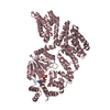
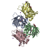
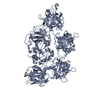
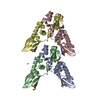
| Details | The biological assembly consists of a crystallographic tetramer made up of molecules A,B,C, and D. |
-Components
| #1: Protein | Mass: 17936.422 Da / Num. of mol.: 4 Source method: isolated from a genetically manipulated source Source: (gene. exp.) Saccharomyces cerevisiae (brewer's yeast)Gene: UBC9 / Plasmid: pGEX / Production host:  Escherichia coli (E. coli) / Strain (production host): BL21(DE3)Gold / References: UniProt: P50623, ubiquitin-protein ligase Escherichia coli (E. coli) / Strain (production host): BL21(DE3)Gold / References: UniProt: P50623, ubiquitin-protein ligase#2: Water | ChemComp-HOH / | Water Mass: 18.015 Da / Num. of mol.: 473 / Source method: isolated from a natural source / Formula: H2O Mass: 18.015 Da / Num. of mol.: 473 / Source method: isolated from a natural source / Formula: H2O |
|---|
-Experimental details
-Experiment
| Experiment | Method: X-RAY DIFFRACTION / Number of used crystals: 1 |
|---|
- Sample preparation
| Crystal | Density Matthews: 2.29 Å3/Da / Density % sol: 46.18 % |
|---|---|
| Crystal grow | Temperature: 291 K / Method: vapor diffusion, hanging drop / pH: 7.5 Details: 18% PEG 6000, 0.1M HEPES, 0.1M NaBr, 5mM DTT, pH 7.5, VAPOR DIFFUSION, HANGING DROP, temperature 291K |
-Data collection
| Diffraction | Mean temperature: 100 K |
|---|---|
| Diffraction source | Source: SYNCHROTRON / Site: NSLS  / Beamline: X25 / Wavelength: 1.1 Å / Beamline: X25 / Wavelength: 1.1 Å |
| Detector | Type: ADSC QUANTUM 315 / Detector: CCD / Date: Jan 30, 2004 |
| Radiation | Protocol: SINGLE WAVELENGTH / Monochromatic (M) / Laue (L): M / Scattering type: x-ray |
| Radiation wavelength | Wavelength: 1.1 Å / Relative weight: 1 |
| Reflection | Resolution: 1.75→25 Å / Num. all: 65526 / Num. obs: 65226 / % possible obs: 99.5 % / Observed criterion σ(F): 0 / Observed criterion σ(I): 0 |
| Reflection shell | Resolution: 1.75→1.81 Å / % possible all: 98.9 |
- Processing
| Software |
| ||||||||||||||||||||
|---|---|---|---|---|---|---|---|---|---|---|---|---|---|---|---|---|---|---|---|---|---|
| Refinement | Method to determine structure: MOLECULAR REPLACEMENT Starting model: PDB ENTRY 1U9A Resolution: 1.75→25 Å / σ(F): 0 / Stereochemistry target values: Engh & Huber
| ||||||||||||||||||||
| Refinement step | Cycle: LAST / Resolution: 1.75→25 Å
| ||||||||||||||||||||
| Xplor file |
|
