 Movie
Movie Controller

[English] 日本語
 Yorodumi
- PDB-2gew: Atomic resolution structure of cholesterol oxidase @ pH 9.0 (Stre... -
+ Open data

- Basic information
| Entry | Database: PDB / ID: 2gew | ||||||
|---|---|---|---|---|---|---|---|
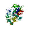
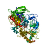
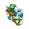
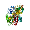
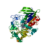
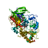
| Title | Atomic resolution structure of cholesterol oxidase @ pH 9.0 (Streptomyces sp. SA-COO) | ||||||
 Components Components | Cholesterol oxidase | ||||||
 Keywords Keywords | OXIDOREDUCTASE / FLAVOENZYME / STEROID METABOLISM / ATOMIC RESOLUTION / HYDROGEN BOND | ||||||
| Function / homology |  Function and homology informationcholesterol oxidase / cholesterol oxidase activity / steroid Delta-isomerase / steroid delta-isomerase activity / cholesterol catabolic process / flavin adenine dinucleotide binding / extracellular region Function and homology informationcholesterol oxidase / cholesterol oxidase activity / steroid Delta-isomerase / steroid delta-isomerase activity / cholesterol catabolic process / flavin adenine dinucleotide binding / extracellular regionSimilarity search - Function | ||||||
| Biological species |  Streptomyces sp. (bacteria) Streptomyces sp. (bacteria) | ||||||
| Method | X-RAY DIFFRACTION / SYNCHROTRON / FOURIER SYNTHESIS / Resolution: 0.97 Å | ||||||
 Authors Authors | Lyubimov, A.Y. / Vrielink, A. | ||||||
 Citation Citation | Journal: Nat.Chem.Biol. / Year: 2006 Title: Atomic resolution crystallography reveals how changes in pH shape the protein microenvironment. Authors: Lyubimov, A.Y. / Lario, P.I. / Moustafa, I. / Vrielink, A. | ||||||
| History |
| ||||||
| Remark 999 | SEQUENCE THE SEQUENCE OF MATURE CHOLESTEROL OXIDASE ENZYME RUNS FROM RESIDUES 43 - 546 IN THE ...SEQUENCE THE SEQUENCE OF MATURE CHOLESTEROL OXIDASE ENZYME RUNS FROM RESIDUES 43 - 546 IN THE SEQUENCE DATABASE. THE NUMBERING OF THE RESIDUES DIFFERS FROM THAT IN THE DATABASE DUE TO THIS DESCREPANCY IN THE SEQUENCE. IN ADDITION, THE NUMBERING HAS BEEN ADJUSTED TO CONFORM TO THAT FOUND IN THE STRUCTURE OF CHOLESTEROL OXIDASE FROM BREVIBACTERIUM STEROLICUM (ACCESSION CODE 3COX). THIS CHANGE IN THE NUMBERING SCHEME FACILITATES EASY COMPARISON OF THE TWO STRUCTURES. |
- Structure visualization
| Structure viewer | Molecule: MolmilJmol/JSmol |
|---|
- Downloads & links
-Download
| PDBx/mmCIF format | 2gew.cif.gz | 342.8 KB | Display | PDBx/mmCIF format |
|---|---|---|---|---|
| PDB format | pdb2gew.ent.gz | 280.8 KB | Display | PDB format |
| PDBx/mmJSON format | 2gew.json.gz | Tree view | PDBx/mmJSON format | |
| Others |  Other downloads Other downloads |
-Validation report
| Arichive directory | https://data.pdbj.org/pub/pdb/validation_reports/ge/2gewftp://data.pdbj.org/pub/pdb/validation_reports/ge/2gew | HTTPS FTP |
|---|
-Related structure data
| Related structure data | 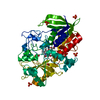 1n4uC  1n4vC  1n4wC  1mxtS S: Starting model for refinement C: citing same article ( |
|---|---|
| Similar structure data |


-Links
 PDBj
PDBj


- Assembly
| Deposited unit | 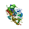
| ||||||||
|---|---|---|---|---|---|---|---|---|---|
| 1 |
| ||||||||
| Unit cell |
|
-Components
| #1: Protein | / CHOD Mass: 54969.676 Da / Num. of mol.: 1 Source method: isolated from a genetically manipulated source Details: FAD COFACTOR NON-COVALENTLY BOUND TO THE ENZYME / Source: (gene. exp.) Streptomyces sp. (bacteria) / Strain: SA-COO / Gene: choA / Plasmid: PCO202 / Production host: Escherichia coli (E. coli) / Strain (production host): BL21(DE3)PLYSS / References: UniProt: P12676, cholesterol oxidase |
|---|---|
| #2: Chemical | ChemComp-SO4 / Sulfate  Mass: 96.063 Da / Num. of mol.: 1 / Source method: obtained synthetically / Formula: SO4 Mass: 96.063 Da / Num. of mol.: 1 / Source method: obtained synthetically / Formula: SO4 |
| #3: Chemical | ChemComp-FAD / Flavin adenine dinucleotide  Mass: 785.550 Da / Num. of mol.: 1 / Source method: obtained synthetically / Formula: C27H33N9O15P2 / Comment: FAD*YM Mass: 785.550 Da / Num. of mol.: 1 / Source method: obtained synthetically / Formula: C27H33N9O15P2 / Comment: FAD*YM |
| #4: Chemical | ChemComp-OXY / Oxygen  Mass: 31.999 Da / Num. of mol.: 1 / Source method: obtained synthetically / Formula: O2 Mass: 31.999 Da / Num. of mol.: 1 / Source method: obtained synthetically / Formula: O2 |
| #5: Water | ChemComp-HOH / Water Mass: 18.015 Da / Num. of mol.: 840 / Source method: isolated from a natural source / Formula: H2O Mass: 18.015 Da / Num. of mol.: 840 / Source method: isolated from a natural source / Formula: H2O |
-Experimental details
-Experiment
| Experiment | Method: X-RAY DIFFRACTION / Number of used crystals: 1 |
|---|
- Sample preparation
| Crystal | Density Matthews: 2.06 Å3/Da / Density % sol: 40.34 % |
|---|---|
| Crystal grow | Temperature: 298 K / Method: vapor diffusion, hanging drop / pH: 9 Details: PEG 8000, AMMONIUM SULFATE, PIPES PH 7.5,VAPOR DIFFUSION, HANGING DROP, temperature 298K, SOAK CONDITION STEP 1: PEG 8000, AMMONIUM SULFATE, HEPES PH 8.0 SOAK CONDITION STEP 2: PEG 8000, ...Details: PEG 8000, AMMONIUM SULFATE, PIPES PH 7.5,VAPOR DIFFUSION, HANGING DROP, temperature 298K, SOAK CONDITION STEP 1: PEG 8000, AMMONIUM SULFATE, HEPES PH 8.0 SOAK CONDITION STEP 2: PEG 8000, AMMONIUM SULFATE, TRICINE PH 8.5 SOAK CONDITION STEP 3: PEG 8000, AMMONIUM SULFATE, CHES PH 9.0, |
-Data collection
| Diffraction | Mean temperature: 100 K |
|---|---|
| Diffraction source | Source: SYNCHROTRON / Site: ALS  / Beamline: 8.2.2 / Wavelength: 0.979 Å / Beamline: 8.2.2 / Wavelength: 0.979 Å |
| Detector | Type: ADSC QUANTUM 315 / Detector: CCD / Date: Jan 22, 2003 |
| Radiation | Monochromator: CRYSTALS SI(111) / Protocol: SINGLE WAVELENGTH / Monochromatic (M) / Laue (L): M / Scattering type: x-ray |
| Radiation wavelength | Wavelength: 0.979 Å / Relative weight: 1 |
| Reflection | Resolution: 0.97→31.3 Å / Num. obs: 243484 / % possible obs: 93 % / Redundancy: 4.5 % / Biso Wilson estimate: 7.8 Å2 / Rmerge(I) obs: 0.075 / Net I/σ(I): 11.8 |
| Reflection shell | Resolution: 0.97→1 Å / Redundancy: 2.2 % / Rmerge(I) obs: 0.51 / Mean I/σ(I) obs: 1.9 / Num. unique all: 20631 / % possible all: 79 |
- Processing
| Software |
| |||||||||||||||||||||||||||||||||
|---|---|---|---|---|---|---|---|---|---|---|---|---|---|---|---|---|---|---|---|---|---|---|---|---|---|---|---|---|---|---|---|---|---|---|
| Refinement | Method to determine structure: FOURIER SYNTHESIS Starting model: 1MXT Resolution: 0.97→31.3 Å / Num. parameters: 46825 / Num. restraintsaints: 130462 / Cross valid method: FREE R / σ(F): 0 / Stereochemistry target values: Engh & Huber
| |||||||||||||||||||||||||||||||||
| Refine analyze | Num. disordered residues: 112 / Occupancy sum hydrogen: 3423.06 / Occupancy sum non hydrogen: 4574.04 | |||||||||||||||||||||||||||||||||
| Refinement step | Cycle: LAST / Resolution: 0.97→31.3 Å
| |||||||||||||||||||||||||||||||||
| Refine LS restraints |
|
