 Movie
Movie Controller


+ Open data

- Basic information
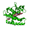
| Entry | Database: PDB / ID: 2gdr | ||||||
|---|---|---|---|---|---|---|---|
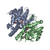
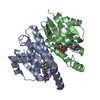
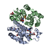
| Title | Crystal structure of a bacterial glutathione transferase | ||||||
 Components Components | glutathione S-transferase | ||||||
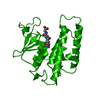
 Keywords Keywords | TRANSFERASE / protein homodimer / each monomer contains two domains / N-term domain is mixed beta sheets and alpha helices / C-term domain is alpha helical | ||||||
| Function / homology |  Function and homology information Function and homology information | ||||||
| Biological species |  Burkholderia xenovorans (bacteria) Burkholderia xenovorans (bacteria) | ||||||
| Method | X-RAY DIFFRACTION / SYNCHROTRON / MOLECULAR REPLACEMENT / Resolution: 2.1 Å | ||||||
 Authors Authors | Tocheva, E.I. / Fortin, P.D. / Eltis, L.D. / Murphy, M.E.P. | ||||||
 Citation Citation | Journal: J.Biol.Chem. / Year: 2006 Title: Structures of Ternary Complexes of BphK, a Bacterial Glutathione S-Transferase That Reductively Dechlorinates Polychlorinated Biphenyl Metabolites. Authors: Tocheva, E.I. / Fortin, P.D. / Eltis, L.D. / Murphy, M.E. | ||||||
| History |
|
- Structure visualization
| Structure viewer | Molecule: MolmilJmol/JSmol |
|---|
- Downloads & links
-Download
| PDBx/mmCIF format | 2gdr.cif.gz | 243.9 KB | Display | PDBx/mmCIF format |
|---|---|---|---|---|
| PDB format | pdb2gdr.ent.gz | 199.7 KB | Display | PDB format |
| PDBx/mmJSON format | 2gdr.json.gz | Tree view | PDBx/mmJSON format | |
| Others |  Other downloads Other downloads |
-Validation report
| Arichive directory | https://data.pdbj.org/pub/pdb/validation_reports/gd/2gdrftp://data.pdbj.org/pub/pdb/validation_reports/gd/2gdr | HTTPS FTP |
|---|
-Related structure data
| Related structure data |  2dsaC  2gdh S: Starting model for refinement C: citing same article ( |
|---|---|
| Similar structure data |


-Links
 PDBj
PDBj

- Assembly
| Deposited unit | 
| ||||||||
|---|---|---|---|---|---|---|---|---|---|
| 1 | 
| ||||||||
| 2 | 
| ||||||||
| 3 | 
| ||||||||
| Unit cell |
|
-Components
| #1: Protein | Mass: 22268.111 Da / Num. of mol.: 6 Source method: isolated from a genetically manipulated source Source: (gene. exp.) Burkholderia xenovorans (bacteria) / Strain: LB400 / Gene: bphK / Production host: Escherichia coli (E. coli)References: GenBank: 27528348, UniProt: Q59721*PLUS, glutathione transferase#2: Chemical | ChemComp-GSH / Glutathione  Mass: 307.323 Da / Num. of mol.: 9 / Source method: obtained synthetically / Formula: C10H17N3O6S Mass: 307.323 Da / Num. of mol.: 9 / Source method: obtained synthetically / Formula: C10H17N3O6S#3: Water | ChemComp-HOH / | Water Mass: 18.015 Da / Num. of mol.: 215 / Source method: isolated from a natural source / Formula: H2O Mass: 18.015 Da / Num. of mol.: 215 / Source method: isolated from a natural source / Formula: H2O |
|---|
-Experimental details
-Experiment
| Experiment | Method: X-RAY DIFFRACTION / Number of used crystals: 1 |
|---|
- Sample preparation
| Crystal | Density Matthews: 3.03 Å3/Da / Density % sol: 59.41 % |
|---|---|
| Crystal grow | Temperature: 323 K / Method: vapor diffusion, hanging drop / pH: 6 Details: 1M K/Na tartrate, 0.1 M MES, pH 6, VAPOR DIFFUSION, HANGING DROP, temperature 323K |
-Data collection
| Diffraction | Mean temperature: 200 K |
|---|---|
| Diffraction source | Source: SYNCHROTRON / Site: SSRL  / Beamline: BL9-1 / Wavelength: 0.97974 Å / Beamline: BL9-1 / Wavelength: 0.97974 Å |
| Detector | Type: ADSC QUANTUM 315 / Detector: CCD / Date: Apr 11, 2005 |
| Radiation | Protocol: SINGLE WAVELENGTH / Monochromatic (M) / Laue (L): M / Scattering type: x-ray |
| Radiation wavelength | Wavelength: 0.97974 Å / Relative weight: 1 |
| Reflection | Resolution: 2.1→50 Å / Num. all: 92159 / Num. obs: 92159 / % possible obs: 97 % / Redundancy: 4.6 % / Rmerge(I) obs: 0.061 |
| Reflection shell | Resolution: 2.1→2.18 Å / Redundancy: 3.4 % / Rmerge(I) obs: 0.384 / % possible all: 95.4 |
- Processing
| Software |
| ||||||||||||||||||||
|---|---|---|---|---|---|---|---|---|---|---|---|---|---|---|---|---|---|---|---|---|---|
| Refinement | Method to determine structure: MOLECULAR REPLACEMENT Starting model: PDB entry 2GDH 2gdh Resolution: 2.1→50 Å / Isotropic thermal model: Isotropic / Stereochemistry target values: Engh & Huber Details: The twinning operator is h,-h-k,-l. The twinning fraction is 0.45.
| ||||||||||||||||||||
| Refinement step | Cycle: LAST / Resolution: 2.1→50 Å
| ||||||||||||||||||||
| LS refinement shell | Resolution: 2.1→2.18 Å / Num. reflection Rfree: 334 |
