 Movie
Movie Controller

[English] 日本語
 Yorodumi
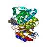
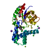
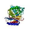
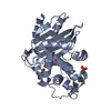
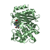
- PDB-2g5x: Crystal structure of lychnin a type 1 Ribosome Inactivating Prote... -
+ Open data

- Basic information
| Entry | Database: PDB / ID: 2g5x | ||||||
|---|---|---|---|---|---|---|---|
| Title | Crystal structure of lychnin a type 1 Ribosome Inactivating Protein (RIP) | ||||||
 Components Components | Ribosome-inactivating protein | ||||||
 Keywords Keywords | HYDROLASE / alpha-beta protein | ||||||
| Function / homology | Ricin (A Subunit), domain 2 / Ricin (A Subunit), domain 2 / Ricin (A subunit); domain 1 / Ricin (A subunit), domain 1 / Few Secondary Structures / Irregular / 3-Layer(aba) Sandwich / Alpha Beta Function and homology information Function and homology information | ||||||
| Biological species |  Lychnis chalcedonica (scarlet lychnis) Lychnis chalcedonica (scarlet lychnis) | ||||||
| Method | X-RAY DIFFRACTION / SYNCHROTRON / MOLECULAR REPLACEMENT / Resolution: 1.7 Å | ||||||
 Authors Authors | Fermani, S. / Falini, G. / Tosi, G. / Ripamonti, A. / Polito, L. / Bolognesi, A. / Stirpe, F. | ||||||
 Citation Citation | Journal: To be Published Title: Crystal structure of lychnin a type 1 Ribosome Inactivating Protein (RIP) Authors: Fermani, S. / Falini, G. / Tosi, G. / Ripamonti, A. / Polito, L. / Bolognesi, A. / Stirpe, F. #1: Journal: Biochim.Biophys.Acta / Year: 1990 Title: Purification and properties of new ribosome-inactivating proteins with RNA N-glycosidase activity Authors: Bolognesi, A. / Barbieri, L. / Abbondanza, A. / Falasca, A.I. / Carnicelli, D. / Battelli, M.G. / Stirpe, F. #2: Journal: ACTA CRYSTALLOGR.,SECT.D / Year: 2003 Title: Crystallization and preliminary X-ray diffraction analysis of two ribosome-inactivating proteins: lychnin and dianthin 30 Authors: Fermani, S. / Falini, G. / Ripamonti, A. / Bolognesi, A. / Polito, L. / Stirpe, F. | ||||||
| History |
|
- Structure visualization
| Structure viewer | Molecule: MolmilJmol/JSmol |
|---|
- Downloads & links
-Download
| PDBx/mmCIF format | 2g5x.cif.gz | 67.2 KB | Display | PDBx/mmCIF format |
|---|---|---|---|---|
| PDB format | pdb2g5x.ent.gz | 48 KB | Display | PDB format |
| PDBx/mmJSON format | 2g5x.json.gz | Tree view | PDBx/mmJSON format | |
| Others |  Other downloads Other downloads |
-Validation report
| Arichive directory | https://data.pdbj.org/pub/pdb/validation_reports/g5/2g5xftp://data.pdbj.org/pub/pdb/validation_reports/g5/2g5x | HTTPS FTP |
|---|
-Related structure data
| Related structure data |  1wuc S: Starting model for refinement |
|---|---|
| Similar structure data |


-Links
 PDBj
PDBj- Assembly
| Deposited unit | 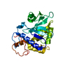
| ||||||||
|---|---|---|---|---|---|---|---|---|---|
| 1 |
| ||||||||
| Unit cell |
| ||||||||
| Details | The protein is a monomer. The asymmetric unit contain one monomer |
-Components
| #1: Protein | / LYCHNIN Mass: 26163.727 Da / Num. of mol.: 1 / Source method: isolated from a natural source / Source: (natural) Lychnis chalcedonica (scarlet lychnis) / Tissue: seedsSeed / References: rRNA N-glycosylase |
|---|---|
| #2: Water | ChemComp-HOH / Water Mass: 18.015 Da / Num. of mol.: 364 / Source method: isolated from a natural source / Formula: H2O Mass: 18.015 Da / Num. of mol.: 364 / Source method: isolated from a natural source / Formula: H2O |
-Experimental details
-Experiment
| Experiment | Method: X-RAY DIFFRACTION / Number of used crystals: 1 |
|---|
- Sample preparation
| Crystal | Density Matthews: 1.97 Å3/Da / Density % sol: 37.66 % |
|---|---|
| Crystal grow | Temperature: 293 K / Method: vapor diffusion, sitting drop / pH: 6.5 Details: 30% PEG 8000, 0.1M sodium phosphate, pH 6.5, VAPOR DIFFUSION, SITTING DROP, temperature 293K |
-Data collection
| Diffraction | Mean temperature: 100 K |
|---|---|
| Diffraction source | Source: SYNCHROTRON / Site: ELETTRA  / Beamline: 5.2R / Wavelength: 1 Å / Beamline: 5.2R / Wavelength: 1 Å |
| Detector | Type: MARRESEARCH / Detector: CCD / Date: Dec 17, 2002 / Details: mirrors |
| Radiation | Monochromator: Si 111 / Protocol: SINGLE WAVELENGTH / Monochromatic (M) / Laue (L): M / Scattering type: x-ray |
| Radiation wavelength | Wavelength: 1 Å / Relative weight: 1 |
| Reflection | Resolution: 1.7→29.1 Å / Num. all: 22021 / Num. obs: 21895 / % possible obs: 51 % / Observed criterion σ(F): 3 / Observed criterion σ(I): -3 / Redundancy: 6.9 % / Biso Wilson estimate: 13.8 Å2 / Rsym value: 0.048 / Net I/σ(I): 39.7 |
| Reflection shell | Resolution: 1.7→1.76 Å / Mean I/σ(I) obs: 14.2 / Num. unique all: 2162 / Rsym value: 0.084 / % possible all: 50.2 |
- Processing
| Software |
| ||||||||||||||||||||||||||||||||||||
|---|---|---|---|---|---|---|---|---|---|---|---|---|---|---|---|---|---|---|---|---|---|---|---|---|---|---|---|---|---|---|---|---|---|---|---|---|---|
| Refinement | Method to determine structure: MOLECULAR REPLACEMENT Starting model: PDB ENTRY 1WUC 1wuc Resolution: 1.7→28.44 Å / Rfactor Rfree error: 0.004 / Data cutoff high absF: 948638.52 / Data cutoff low absF: 0 / Isotropic thermal model: RESTRAINED / Cross valid method: THROUGHOUT / σ(F): 0 / Stereochemistry target values: Engh & Huber
| ||||||||||||||||||||||||||||||||||||
| Solvent computation | Solvent model: FLAT MODEL / Bsol: 80.4733 Å2 / ksol: 0.460368 e/Å3 | ||||||||||||||||||||||||||||||||||||
| Displacement parameters | Biso mean: 13.6 Å2
| ||||||||||||||||||||||||||||||||||||
| Refine analyze |
| ||||||||||||||||||||||||||||||||||||
| Refinement step | Cycle: LAST / Resolution: 1.7→28.44 Å
| ||||||||||||||||||||||||||||||||||||
| Refine LS restraints |
| ||||||||||||||||||||||||||||||||||||
| LS refinement shell | Resolution: 1.7→1.81 Å / Rfactor Rfree error: 0.013 / Total num. of bins used: 6
| ||||||||||||||||||||||||||||||||||||
| Xplor file |
|
