 Movie
Movie Controller

[English] 日本語
 Yorodumi
- PDB-2g3n: Crystal structure of the Sulfolobus solfataricus alpha-glucosidas... -
+ Open data

- Basic information
| Entry | Database: PDB / ID: 2g3n | ||||||
|---|---|---|---|---|---|---|---|
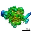
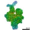
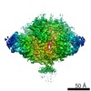
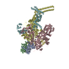
| Title | Crystal structure of the Sulfolobus solfataricus alpha-glucosidase MalA in complex with beta-octyl-glucopyranoside | ||||||
 Components Components | Alpha-glucosidase | ||||||
 Keywords Keywords | HYDROLASE / alpha-glucosidase / enzyme-carbohydrate complex / glycoside hydrolase family 31 / multidomain protein / (beta/alpha)8 barrel / retaining mechanism | ||||||
| Function / homology |  Function and homology informationalpha-glucosidase / maltose alpha-glucosidase activity / N-glycan processing / hydrolase activity, hydrolyzing O-glycosyl compounds / carbohydrate binding / cytoplasm Function and homology informationalpha-glucosidase / maltose alpha-glucosidase activity / N-glycan processing / hydrolase activity, hydrolyzing O-glycosyl compounds / carbohydrate binding / cytoplasmSimilarity search - Function | ||||||
| Biological species |   Sulfolobus solfataricus (archaea) Sulfolobus solfataricus (archaea) | ||||||
| Method | X-RAY DIFFRACTION / SYNCHROTRON / MOLECULAR REPLACEMENT / Resolution: 2.55 Å | ||||||
 Authors Authors | Ernst, H.A. / Lo Leggio, L. / Willemoes, M. / Leonard, G. / Blum, P. / Larsen, S. | ||||||
 Citation Citation | Journal: J.Mol.Biol. / Year: 2006 Title: Structure of the Sulfolobus solfataricus alpha-Glucosidase: Implications for Domain Conservation and Substrate Recognition in GH31. Authors: Ernst, H.A. / Lo Leggio, L. / Willemoes, M. / Leonard, G. / Blum, P. / Larsen, S. #1: Journal: Acta Crystallogr.,Sect.F / Year: 2006Title: Characterization of different crystal forms of the alpha-glucosidase MalA from Sulfolobus solfataricus Authors: Ernst, H.A. / Willemoes, M. / Lo Leggio, L. / Leonard, G. / Blum, P. / Larsen, S. | ||||||
| History |
|
- Structure visualization
| Structure viewer | Molecule: MolmilJmol/JSmol |
|---|
- Downloads & links
-Download
| PDBx/mmCIF format | 2g3n.cif.gz | 830.9 KB | Display | PDBx/mmCIF format |
|---|---|---|---|---|
| PDB format | pdb2g3n.ent.gz | 685.2 KB | Display | PDB format |
| PDBx/mmJSON format | 2g3n.json.gz | Tree view | PDBx/mmJSON format | |
| Others |  Other downloads Other downloads |
-Validation report
| Arichive directory | https://data.pdbj.org/pub/pdb/validation_reports/g3/2g3nftp://data.pdbj.org/pub/pdb/validation_reports/g3/2g3n | HTTPS FTP |
|---|
-Related structure data
| Related structure data | 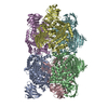 2g3mSC S: Starting model for refinement C: citing same article ( |
|---|---|
| Similar structure data |


-Links
 PDBj
PDBj


- Assembly
| Deposited unit | 
| ||||||||
|---|---|---|---|---|---|---|---|---|---|
| 1 |
| ||||||||
| Unit cell |
|
-Components
| #1: Protein | / Maltase Mass: 80580.195 Da / Num. of mol.: 6 Source method: isolated from a genetically manipulated source Source: (gene. exp.) Sulfolobus solfataricus (archaea) / Strain: 98-2 / Gene: malA / Plasmid: pMW800 / Production host:  Escherichia coli (E. coli) / Strain (production host): NF1830 Escherichia coli (E. coli) / Strain (production host): NF1830References: UniProt: O59645, UniProt: P0CD66*PLUS, alpha-glucosidase#2: Sugar | ChemComp-BOG / Octyl glucoside  Type: D-saccharide / Mass: 292.369 Da / Num. of mol.: 6 Type: D-saccharide / Mass: 292.369 Da / Num. of mol.: 6Source method: isolated from a genetically manipulated source Formula: C14H28O6 / Comment: detergent*YM #3: Water | ChemComp-HOH / | Water Mass: 18.015 Da / Num. of mol.: 615 / Source method: isolated from a natural source / Formula: H2O Mass: 18.015 Da / Num. of mol.: 615 / Source method: isolated from a natural source / Formula: H2O |
|---|
-Experimental details
-Experiment
| Experiment | Method: X-RAY DIFFRACTION / Number of used crystals: 1 |
|---|
- Sample preparation
| Crystal | Density Matthews: 2.46 Å3/Da / Density % sol: 49.94 % |
|---|---|
| Crystal grow | Temperature: 298 K / Method: vapor diffusion, hanging drop / pH: 4.2 Details: 10 % PEG 4000, 0.2 M sodium citrate, 0.1 M sodium acetate, 0.5 % beta-octyl-glucopyranoside, microseeding, pH 4.2, VAPOR DIFFUSION, HANGING DROP, temperature 298K |
-Data collection
| Diffraction | Mean temperature: 100 K |
|---|---|
| Diffraction source | Source: SYNCHROTRON / Site: ESRF  / Beamline: ID14-1 / Wavelength: 0.934 Å / Beamline: ID14-1 / Wavelength: 0.934 Å |
| Detector | Type: ADSC QUANTUM 4 / Detector: CCD / Date: Nov 27, 2004 |
| Radiation | Protocol: SINGLE WAVELENGTH / Monochromatic (M) / Laue (L): M / Scattering type: x-ray |
| Radiation wavelength | Wavelength: 0.934 Å / Relative weight: 1 |
| Reflection | Resolution: 2.5→24.2 Å / Num. obs: 154885 / % possible obs: 95.2 % / Observed criterion σ(F): 0 / Observed criterion σ(I): 0 / Redundancy: 2.6 % / Biso Wilson estimate: 34.5 Å2 / Rmerge(I) obs: 0.065 / Net I/σ(I): 11.67 |
| Reflection shell | Resolution: 2.5→2.63 Å / Redundancy: 2.3 % / Rmerge(I) obs: 0.29 / Mean I/σ(I) obs: 5.3 |
-Phasing
| Phasing MR | Rfactor: 0.391 / Cor.coef. Fo:Fc: 0.643
|
|---|
- Processing
| Software |
| ||||||||||||||||||||||||||||||||||||||||||||||||||||||||||||||||||||||||||||||||
|---|---|---|---|---|---|---|---|---|---|---|---|---|---|---|---|---|---|---|---|---|---|---|---|---|---|---|---|---|---|---|---|---|---|---|---|---|---|---|---|---|---|---|---|---|---|---|---|---|---|---|---|---|---|---|---|---|---|---|---|---|---|---|---|---|---|---|---|---|---|---|---|---|---|---|---|---|---|---|---|---|---|
| Refinement | Method to determine structure: MOLECULAR REPLACEMENT Starting model: A trimmed version of the trimer in PDB code 2G3M Resolution: 2.55→24.16 Å / Rfactor Rfree error: 0.002 / Data cutoff high absF: 3189066.23 / Data cutoff low absF: 0 / Isotropic thermal model: RESTRAINED / Cross valid method: THROUGHOUT / σ(F): 0 / Stereochemistry target values: maximum likelihood
| ||||||||||||||||||||||||||||||||||||||||||||||||||||||||||||||||||||||||||||||||
| Solvent computation | Solvent model: FLAT MODEL / Bsol: 33.366 Å2 / ksol: 0.382443 e/Å3 | ||||||||||||||||||||||||||||||||||||||||||||||||||||||||||||||||||||||||||||||||
| Displacement parameters | Biso mean: 43.2 Å2
| ||||||||||||||||||||||||||||||||||||||||||||||||||||||||||||||||||||||||||||||||
| Refine analyze |
| ||||||||||||||||||||||||||||||||||||||||||||||||||||||||||||||||||||||||||||||||
| Refinement step | Cycle: LAST / Resolution: 2.55→24.16 Å
| ||||||||||||||||||||||||||||||||||||||||||||||||||||||||||||||||||||||||||||||||
| Refine LS restraints |
| ||||||||||||||||||||||||||||||||||||||||||||||||||||||||||||||||||||||||||||||||
| Refine LS restraints NCS | NCS model details: RESTRAINED | ||||||||||||||||||||||||||||||||||||||||||||||||||||||||||||||||||||||||||||||||
| LS refinement shell | Resolution: 2.55→2.71 Å / Rfactor Rfree error: 0.009 / Total num. of bins used: 6
| ||||||||||||||||||||||||||||||||||||||||||||||||||||||||||||||||||||||||||||||||
| Xplor file |
|
