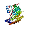
 Movie
Movie Controller


+ Open data

- Basic information
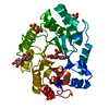
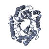
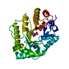
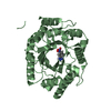
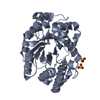
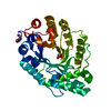
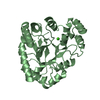
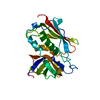
| Entry | Database: PDB / ID: 2g3i | ||||||
|---|---|---|---|---|---|---|---|
| Title | Structure of S.olivaceoviridis xylanase Q88A/R275A mutant | ||||||
 Components Components | Xylanase | ||||||
 Keywords Keywords | HYDROLASE / glycoside hydrolase | ||||||
| Function / homology |  Function and homology informationendo-1,4-beta-xylanase activity / endo-1,4-beta-xylanase / polysaccharide catabolic process Function and homology informationendo-1,4-beta-xylanase activity / endo-1,4-beta-xylanase / polysaccharide catabolic processSimilarity search - Function | ||||||
| Biological species |  Streptomyces olivaceoviridis (bacteria) Streptomyces olivaceoviridis (bacteria) | ||||||
| Method | X-RAY DIFFRACTION / SYNCHROTRON / FOURIER SYNTHESIS / Resolution: 2.1 Å | ||||||
 Authors Authors | Diertavitian, S. / Kaneko, S. / Fujimoto, Z. / Kuno, A. / Johansson, E. / Lo Leggio, L. | ||||||
 Citation Citation | Journal: PROCESS BIOCHEM / Year: 2012 Title: Structure-based engineering of glucose specificity in a family 10 xylanase from Streptomyces olivaceoviridis E-86 Authors: Ichinose, H. / Diertavitian, S. / Fujimoto, Z. / Kuno, A. / Leggio, L.L. / Kaneko, S. | ||||||
| History |
|
- Structure visualization
| Structure viewer | Molecule: MolmilJmol/JSmol |
|---|
- Downloads & links
-Download
| PDBx/mmCIF format | 2g3i.cif.gz | 77.9 KB | Display | PDBx/mmCIF format |
|---|---|---|---|---|
| PDB format | pdb2g3i.ent.gz | 57.8 KB | Display | PDB format |
| PDBx/mmJSON format | 2g3i.json.gz | Tree view | PDBx/mmJSON format | |
| Others |  Other downloads Other downloads |
-Validation report
| Arichive directory | https://data.pdbj.org/pub/pdb/validation_reports/g3/2g3iftp://data.pdbj.org/pub/pdb/validation_reports/g3/2g3i | HTTPS FTP |
|---|
-Related structure data


-Links
 PDBj
PDBj


- Assembly
| Deposited unit | 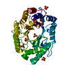
| |||||||||
|---|---|---|---|---|---|---|---|---|---|---|
| 1 |
| |||||||||
| Unit cell |
| |||||||||
| Components on special symmetry positions |
|
-Components
| #1: Protein | Mass: 34034.383 Da / Num. of mol.: 1 / Fragment: catalytic domain / Mutation: Q88A/R275A Source method: isolated from a genetically manipulated source Source: (gene. exp.) Streptomyces olivaceoviridis (bacteria)Strain: E-86 / Gene: Xylanase / Plasmid: PET28A (Novagen) / Species (production host): Escherichia coli / Production host: Escherichia coli BL21(DE3) (bacteria) / Strain (production host): BL21 (DE3) / References: UniProt: Q7SI98, endo-1,4-beta-xylanase | ||
|---|---|---|---|
| #2: Chemical | ChemComp-PO4 / Phosphate  Mass: 94.971 Da / Num. of mol.: 7 / Source method: obtained synthetically / Formula: PO4 Mass: 94.971 Da / Num. of mol.: 7 / Source method: obtained synthetically / Formula: PO4#3: Water | ChemComp-HOH / | Water Mass: 18.015 Da / Num. of mol.: 233 / Source method: isolated from a natural source / Formula: H2O Mass: 18.015 Da / Num. of mol.: 233 / Source method: isolated from a natural source / Formula: H2O |
-Experimental details
-Experiment
| Experiment | Method: X-RAY DIFFRACTION / Number of used crystals: 1 |
|---|
- Sample preparation
| Crystal | Density Matthews: 3.4 Å3/Da / Density % sol: 63.85 % |
|---|---|
| Crystal grow | Temperature: 294 K / Method: vapor diffusion, hanging drop / pH: 8.5 Details: 2.1M ammonium dehydrogen phosphate, 0.1M Tris pH 8.5, VAPOR DIFFUSION, HANGING DROP, temperature 294K |
-Data collection
| Diffraction | Mean temperature: 100 K |
|---|---|
| Diffraction source | Source: SYNCHROTRON / Site: ESRF  / Beamline: ID29 / Wavelength: 0.999 Å / Beamline: ID29 / Wavelength: 0.999 Å |
| Detector | Type: MARRESEARCH / Detector: CCD / Date: Apr 17, 2003 |
| Radiation | Protocol: SINGLE WAVELENGTH / Monochromatic (M) / Laue (L): M / Scattering type: x-ray |
| Radiation wavelength | Wavelength: 0.999 Å / Relative weight: 1 |
| Reflection | Resolution: 2.1→30 Å / Num. obs: 25881 / % possible obs: 99.5 % / Redundancy: 8.4 % / Rsym value: 0.179 / Net I/σ(I): 13.3 |
| Reflection shell | Resolution: 2.1→2.2 Å / Redundancy: 5 % / Mean I/σ(I) obs: 4.2 / Rsym value: 0.511 / % possible all: 99.1 |
- Processing
| Software |
| ||||||||||||||||||||||||||||||||||||||||||||||||||||||||||||||||||||||||||||||||||||||||||||||||||||||||||||||||||||||||
|---|---|---|---|---|---|---|---|---|---|---|---|---|---|---|---|---|---|---|---|---|---|---|---|---|---|---|---|---|---|---|---|---|---|---|---|---|---|---|---|---|---|---|---|---|---|---|---|---|---|---|---|---|---|---|---|---|---|---|---|---|---|---|---|---|---|---|---|---|---|---|---|---|---|---|---|---|---|---|---|---|---|---|---|---|---|---|---|---|---|---|---|---|---|---|---|---|---|---|---|---|---|---|---|---|---|---|---|---|---|---|---|---|---|---|---|---|---|---|---|---|---|
| Refinement | Method to determine structure: FOURIER SYNTHESIS Starting model: same protein in different crystal form Resolution: 2.1→19.88 Å / Cor.coef. Fo:Fc: 0.953 / Cor.coef. Fo:Fc free: 0.929 / SU B: 3.249 / SU ML: 0.087 / Cross valid method: THROUGHOUT / σ(F): 0 / ESU R: 0.152 / ESU R Free: 0.14 / Stereochemistry target values: MAXIMUM LIKELIHOOD / Details: HYDROGENS HAVE BEEN ADDED IN THE RIDING POSITIONS
| ||||||||||||||||||||||||||||||||||||||||||||||||||||||||||||||||||||||||||||||||||||||||||||||||||||||||||||||||||||||||
| Solvent computation | Ion probe radii: 0.8 Å / Shrinkage radii: 0.8 Å / VDW probe radii: 1.2 Å / Solvent model: MASK | ||||||||||||||||||||||||||||||||||||||||||||||||||||||||||||||||||||||||||||||||||||||||||||||||||||||||||||||||||||||||
| Displacement parameters | Biso mean: 12.607 Å2
| ||||||||||||||||||||||||||||||||||||||||||||||||||||||||||||||||||||||||||||||||||||||||||||||||||||||||||||||||||||||||
| Refinement step | Cycle: LAST / Resolution: 2.1→19.88 Å
| ||||||||||||||||||||||||||||||||||||||||||||||||||||||||||||||||||||||||||||||||||||||||||||||||||||||||||||||||||||||||
| Refine LS restraints |
| ||||||||||||||||||||||||||||||||||||||||||||||||||||||||||||||||||||||||||||||||||||||||||||||||||||||||||||||||||||||||
| LS refinement shell | Resolution: 2.1→2.154 Å / Total num. of bins used: 20
|
