| Entry | Database: PDB / ID: 2fyf
|
|---|
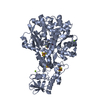
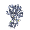
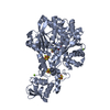
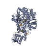
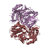
| Title | Structure of a putative phosphoserine aminotransferase from Mycobacterium Tuberculosis |
|---|
 Components Components | phosphoserine aminotransferase Phosphoserine transaminase Phosphoserine transaminase |
|---|
 Keywords Keywords | TRANSFERASE / PLP-dependent enzyme / dimer / Structural Genomics / Mycobacterium Tuberculosis Structural Proteomics Project / XMTB |
|---|
| Function / homology |  Function and homology information Function and homology information
glycine biosynthetic process, by transamination of glyoxylate / serine-pyruvate transaminase activity / phosphoserine transaminase / O-phospho-L-serine:2-oxoglutarate aminotransferase activity / alanine-glyoxylate transaminase activity / pyridoxine biosynthetic process / L-serine biosynthetic process / peroxisome / pyridoxal phosphate binding / extracellular region ...glycine biosynthetic process, by transamination of glyoxylate / serine-pyruvate transaminase activity / phosphoserine transaminase / O-phospho-L-serine:2-oxoglutarate aminotransferase activity / alanine-glyoxylate transaminase activity / pyridoxine biosynthetic process / L-serine biosynthetic process / peroxisome / pyridoxal phosphate binding / extracellular region / plasma membrane / cytoplasmSimilarity search - Function Phosphoserine aminotransferase, Mycobacterial-type / Phosphoserine aminotransferase / Aminotransferase class V domain / Aminotransferase class-V / Aspartate Aminotransferase, domain 1 / Aspartate Aminotransferase, domain 1 / Aspartate Aminotransferase; domain 2 / Type I PLP-dependent aspartate aminotransferase-like (Major domain) / Pyridoxal phosphate-dependent transferase, small domain / Pyridoxal phosphate-dependent transferase, major domain ...Phosphoserine aminotransferase, Mycobacterial-type / Phosphoserine aminotransferase / Aminotransferase class V domain / Aminotransferase class-V / Aspartate Aminotransferase, domain 1 / Aspartate Aminotransferase, domain 1 / Aspartate Aminotransferase; domain 2 / Type I PLP-dependent aspartate aminotransferase-like (Major domain) / Pyridoxal phosphate-dependent transferase, small domain / Pyridoxal phosphate-dependent transferase, major domain / Pyridoxal phosphate-dependent transferase / Alpha-Beta Complex / 3-Layer(aba) Sandwich / Alpha BetaSimilarity search - Domain/homology |
|---|
| Biological species |   Mycobacterium tuberculosis (bacteria) Mycobacterium tuberculosis (bacteria) |
|---|
| Method | X-RAY DIFFRACTION / SYNCHROTRON / MIR / MIRAS / Resolution: 1.5 Å |
|---|
 Authors Authors | Coulibaly, F. / Lassalle, E. / Baker, E.N. / Mycobacterium Tuberculosis Structural Proteomics Project (XMTB) |
|---|
 Citation Citation | Journal: Acta Crystallogr.,Sect.D / Year: 2012
Title: Structure of phosphoserine aminotransferase from Mycobacterium tuberculosis.
Authors: Coulibaly, F. / Lassalle, E. / Baker, H.M. / Baker, E.N. |
|---|
| History | | Deposition | Feb 7, 2006 | Deposition site: RCSB / Processing site: RCSB |
|---|
| Revision 1.0 | Jan 16, 2007 | Provider: repository / Type: Initial release |
|---|
| Revision 1.1 | May 1, 2008 | Group: Version format compliance |
|---|
| Revision 1.2 | Jul 13, 2011 | Group: Advisory / Source and taxonomy / Version format compliance |
|---|
| Revision 1.3 | Jun 13, 2012 | Group: Database references |
|---|
| Revision 1.4 | Oct 18, 2017 | Group: Advisory / Refinement description / Category: pdbx_unobs_or_zero_occ_atoms / software |
|---|
| Revision 1.5 | Feb 14, 2024 | Group: Advisory / Data collection ...Advisory / Data collection / Database references / Derived calculations
Category: chem_comp_atom / chem_comp_bond ...chem_comp_atom / chem_comp_bond / database_2 / pdbx_struct_conn_angle / pdbx_unobs_or_zero_occ_atoms / struct_conn / struct_ref_seq_dif / struct_site
Item: _database_2.pdbx_DOI / _database_2.pdbx_database_accession ..._database_2.pdbx_DOI / _database_2.pdbx_database_accession / _pdbx_struct_conn_angle.ptnr1_auth_comp_id / _pdbx_struct_conn_angle.ptnr1_auth_seq_id / _pdbx_struct_conn_angle.ptnr1_label_asym_id / _pdbx_struct_conn_angle.ptnr1_label_atom_id / _pdbx_struct_conn_angle.ptnr1_label_comp_id / _pdbx_struct_conn_angle.ptnr1_label_seq_id / _pdbx_struct_conn_angle.ptnr3_auth_comp_id / _pdbx_struct_conn_angle.ptnr3_auth_seq_id / _pdbx_struct_conn_angle.ptnr3_label_asym_id / _pdbx_struct_conn_angle.ptnr3_label_atom_id / _pdbx_struct_conn_angle.ptnr3_label_comp_id / _pdbx_struct_conn_angle.ptnr3_label_seq_id / _pdbx_struct_conn_angle.value / _struct_conn.pdbx_dist_value / _struct_conn.pdbx_ptnr1_label_alt_id / _struct_conn.ptnr1_auth_comp_id / _struct_conn.ptnr1_auth_seq_id / _struct_conn.ptnr1_label_asym_id / _struct_conn.ptnr1_label_atom_id / _struct_conn.ptnr1_label_comp_id / _struct_conn.ptnr1_label_seq_id / _struct_conn.ptnr2_auth_comp_id / _struct_conn.ptnr2_auth_seq_id / _struct_conn.ptnr2_label_asym_id / _struct_conn.ptnr2_label_atom_id / _struct_conn.ptnr2_label_comp_id / _struct_conn.ptnr2_label_seq_id / _struct_ref_seq_dif.details / _struct_site.pdbx_auth_asym_id / _struct_site.pdbx_auth_comp_id / _struct_site.pdbx_auth_seq_id |
|---|
|
|---|
 Movie
Movie Controller

 Yorodumi
 Open data
 Basic information
 Structure visualization
 Downloads & links
 Other downloads


 PDBj
PDBj Assembly
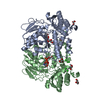


 Mass: 96.063 Da / Num. of mol.: 2 / Source method: obtained synthetically / Formula: SO4
 Mass: 336.890 Da / Num. of mol.: 6 / Source method: obtained synthetically / Formula: Cl4Pt
 Mass: 247.142 Da / Num. of mol.: 2 / Source method: obtained synthetically / Formula: C8H10NO6P
 Mass: 92.094 Da / Num. of mol.: 7 / Source method: obtained synthetically / Formula: C3H8O3
 Sample preparation
 / Beamline: ID29 / Wavelength: 1.07198 Å