 Movie
Movie Controller

[English] 日本語
 Yorodumi
- PDB-2fy6: Structure of the N-terminal domain of Neisseria meningitidis PilB -
+ Open data

- Basic information
| Entry | Database: PDB / ID: 2fy6 | ||||||
|---|---|---|---|---|---|---|---|
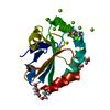
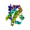
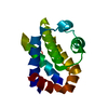
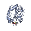
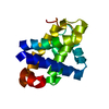
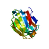
| Title | Structure of the N-terminal domain of Neisseria meningitidis PilB | ||||||
 Components Components | Peptide methionine sulfoxide reductase msrA/msrB | ||||||
 Keywords Keywords |  OXIDOREDUCTASE / PILB / Cytochrome maturation protein / Thioredoxin / Methionine sulfoxide reductase OXIDOREDUCTASE / PILB / Cytochrome maturation protein / Thioredoxin / Methionine sulfoxide reductase | ||||||
| Function / homology |  Function and homology informationL-methionine:thioredoxin-disulfide S-oxidoreductase activity / peptide-methionine (R)-S-oxide reductase / peptide-methionine (R)-S-oxide reductase activity / peptide-methionine (S)-S-oxide reductase / peptide-methionine (S)-S-oxide reductase activity / protein repair / protein modification process / response to oxidative stress Function and homology informationL-methionine:thioredoxin-disulfide S-oxidoreductase activity / peptide-methionine (R)-S-oxide reductase / peptide-methionine (R)-S-oxide reductase activity / peptide-methionine (S)-S-oxide reductase / peptide-methionine (S)-S-oxide reductase activity / protein repair / protein modification process / response to oxidative stressSimilarity search - Function | ||||||
| Biological species |  Neisseria meningitidis (bacteria) Neisseria meningitidis (bacteria) | ||||||
| Method | X-RAY DIFFRACTION / SYNCHROTRON / SAD / Resolution: 1.9 Å | ||||||
 Authors Authors | Ranaivoson, F.M. / Kauffmann, B. / Neiers, F. / Boschi-Muller, S. / Branlant, G. / Favier, F. | ||||||
 Citation Citation | Journal: J.Mol.Biol. / Year: 2006 Title: The X-ray Structure of the N-terminal Domain of PILB from Neisseria meningitidis Reveals a Thioredoxin-fold Authors: Ranaivoson, F.M. / Kauffmann, B. / Neiers, F. / Wu, J. / Boschi-Muller, S. / Panjikar, S. / Aubry, A. / Branlant, G. / Favier, F. | ||||||
| History |
|
- Structure visualization
| Structure viewer | Molecule: MolmilJmol/JSmol |
|---|
- Downloads & links
-Download
| PDBx/mmCIF format | 2fy6.cif.gz | 44 KB | Display | PDBx/mmCIF format |
|---|---|---|---|---|
| PDB format | pdb2fy6.ent.gz | 29.9 KB | Display | PDB format |
| PDBx/mmJSON format | 2fy6.json.gz | Tree view | PDBx/mmJSON format | |
| Others |  Other downloads Other downloads |
-Validation report
| Arichive directory | https://data.pdbj.org/pub/pdb/validation_reports/fy/2fy6ftp://data.pdbj.org/pub/pdb/validation_reports/fy/2fy6 | HTTPS FTP |
|---|
-Related structure data
| Similar structure data |
|---|


-Links
 PDBj
PDBj- Assembly
| Deposited unit | 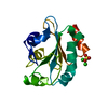
| ||||||||
|---|---|---|---|---|---|---|---|---|---|
| 1 |
| ||||||||
| Unit cell |
|
-Components
| #1: Protein | Mass: 15716.895 Da / Num. of mol.: 1 / Fragment: thioredoxin-like domain Source method: isolated from a genetically manipulated source Source: (gene. exp.) Neisseria meningitidis (bacteria) / Strain: Z2491 / Gene: msrAB, pilB / Plasmid: pET24c / Species (production host): Escherichia coli / Production host: Escherichia coli BL21 (bacteria) / Strain (production host): BL21 / References: UniProt: Q9JWM8, EC: 1.8.4.6 | ||||
|---|---|---|---|---|---|
| #2: Chemical | Chloride  Mass: 35.453 Da / Num. of mol.: 2 / Source method: obtained synthetically / Formula: Cl Mass: 35.453 Da / Num. of mol.: 2 / Source method: obtained synthetically / Formula: Cl#3: Chemical | ChemComp-SO4 / | Sulfate  Mass: 96.063 Da / Num. of mol.: 1 / Source method: obtained synthetically / Formula: SO4 Mass: 96.063 Da / Num. of mol.: 1 / Source method: obtained synthetically / Formula: SO4#4: Water | ChemComp-HOH / | Water Mass: 18.015 Da / Num. of mol.: 154 / Source method: isolated from a natural source / Formula: H2O Mass: 18.015 Da / Num. of mol.: 154 / Source method: isolated from a natural source / Formula: H2O |
-Experimental details
-Experiment
| Experiment | Method: X-RAY DIFFRACTION / Number of used crystals: 1 |
|---|
- Sample preparation
| Crystal | Density Matthews: 2.24 Å3/Da / Density % sol: 45.08 % |
|---|---|
| Crystal grow | Temperature: 293 K / Method: microbatch / pH: 7.5 Details: 3.5M Ammonium Sulfate, Tris-HCl 50mM pH 8.0, 6mg/mL protein, pH 7.5, microbatch, temperature 293K |
-Data collection
| Diffraction | Mean temperature: 100 K |
|---|---|
| Diffraction source | Source: SYNCHROTRON / Site: EMBL/DESY, HAMBURG  / Beamline: X11 / Wavelength: 0.81 Å / Beamline: X11 / Wavelength: 0.81 Å |
| Detector | Type: MARRESEARCH / Detector: CCD / Date: Dec 15, 2003 / Details: Bent mirror |
| Radiation | Monochromator: Triangular / Protocol: SINGLE WAVELENGTH / Monochromatic (M) / Laue (L): M / Scattering type: x-ray |
| Radiation wavelength | Wavelength: 0.81 Å / Relative weight: 1 |
| Reflection | Resolution: 1.9→30 Å / Num. obs: 11090 / % possible obs: 99.9 % / Observed criterion σ(F): 0 / Observed criterion σ(I): 0 / Redundancy: 3.7 % / Biso Wilson estimate: 17.5 Å2 / Rmerge(I) obs: 0.084 / Χ2: 0.537 / Net I/σ(I): 6.9 |
| Reflection shell | Resolution: 1.9→1.97 Å / % possible obs: 100 % / Rmerge(I) obs: 0.405 / Mean I/σ(I) obs: 2.3 / Num. unique all: 1088 / Num. unique obs: 1088 / Χ2: 0.288 / % possible all: 100 |
- Processing
| Software |
| ||||||||||||||||||||||||||||||||||||
|---|---|---|---|---|---|---|---|---|---|---|---|---|---|---|---|---|---|---|---|---|---|---|---|---|---|---|---|---|---|---|---|---|---|---|---|---|---|
| Refinement | Method to determine structure: SAD / Resolution: 1.9→18.62 Å / Rfactor Rfree error: 0.007 / FOM work R set: 0.868 / Data cutoff high absF: 585795.812 / Data cutoff low absF: 0 / Isotropic thermal model: RESTRAINED / Cross valid method: THROUGHOUT / σ(F): 0 / Stereochemistry target values: Engh & Huber
| ||||||||||||||||||||||||||||||||||||
| Solvent computation | Solvent model: FLAT MODEL / Bsol: 49.635 Å2 / ksol: 0.36 e/Å3 | ||||||||||||||||||||||||||||||||||||
| Displacement parameters | Biso mean: 20.5 Å2
| ||||||||||||||||||||||||||||||||||||
| Refine analyze |
| ||||||||||||||||||||||||||||||||||||
| Refinement step | Cycle: LAST / Resolution: 1.9→18.62 Å
| ||||||||||||||||||||||||||||||||||||
| Refine LS restraints |
| ||||||||||||||||||||||||||||||||||||
| LS refinement shell | Resolution: 1.9→2.02 Å / Rfactor Rfree error: 0.02 / Total num. of bins used: 6
| ||||||||||||||||||||||||||||||||||||
| Xplor file |
|
