 Movie
Movie Controller

[English] 日本語
 Yorodumi
- PDB-2fwa: Structure of PurE (N5-carboxyaminoimidazole ribonucleotide mutase... -
+ Open data

- Basic information
| Entry | Database: PDB / ID: 2fwa | ||||||
|---|---|---|---|---|---|---|---|
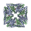
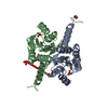
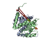
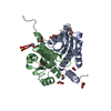
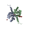
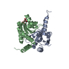
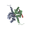
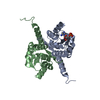
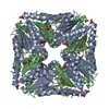
| Title | Structure of PurE (N5-carboxyaminoimidazole ribonucleotide mutase) H89N from the acidophilic bacterium Acetobacter aceti, at pH 7 | ||||||
 Components Components | N5-carboxyaminoimidazole ribonucleotide mutase | ||||||
 Keywords Keywords |  LYASE / acidophile / PurE / purine biosynthesis LYASE / acidophile / PurE / purine biosynthesis | ||||||
| Function / homology |  Function and homology information5-(carboxyamino)imidazole ribonucleotide mutase / 5-(carboxyamino)imidazole ribonucleotide mutase activity / 'de novo' IMP biosynthetic process Function and homology information5-(carboxyamino)imidazole ribonucleotide mutase / 5-(carboxyamino)imidazole ribonucleotide mutase activity / 'de novo' IMP biosynthetic processSimilarity search - Function | ||||||
| Biological species |  Acetobacter aceti (bacteria) Acetobacter aceti (bacteria) | ||||||
| Method | X-RAY DIFFRACTION / FOURIER SYNTHESIS / Resolution: 1.9 Å | ||||||
 Authors Authors | Starks, C.M. / Kappock, T.J. | ||||||
 Citation Citation | Journal: Biochemistry / Year: 2006 Title: Biochemical and Structural Studies of N(5)-Carboxyaminoimidazole Ribonucleotide Mutase from the Acidophilic Bacterium Acetobacter aceti. Authors: Constantine, C.Z. / Starks, C.M. / Mill, C.P. / Ransome, A.E. / Karpowicz, S.J. / Francois, J.A. / Goodman, R.A. / Kappock, T.J. | ||||||
| History |
|
- Structure visualization
| Structure viewer | Molecule: MolmilJmol/JSmol |
|---|
- Downloads & links
-Download
| PDBx/mmCIF format | 2fwa.cif.gz | 75.7 KB | Display | PDBx/mmCIF format |
|---|---|---|---|---|
| PDB format | pdb2fwa.ent.gz | 56.8 KB | Display | PDB format |
| PDBx/mmJSON format | 2fwa.json.gz | Tree view | PDBx/mmJSON format | |
| Others |  Other downloads Other downloads |
-Validation report
| Arichive directory | https://data.pdbj.org/pub/pdb/validation_reports/fw/2fwaftp://data.pdbj.org/pub/pdb/validation_reports/fw/2fwa | HTTPS FTP |
|---|
-Related structure data
| Related structure data | 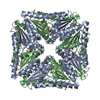 2fw1C  2fw6C 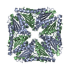 2fw7C 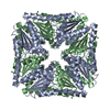 2fw8C 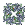 2fw9C 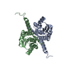 2fwbC  2fwiC 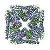 2fwjC 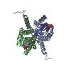 2fwpC 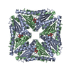 1u11S S: Starting model for refinement C: citing same article ( |
|---|---|
| Similar structure data |

-Links
 PDBj
PDBj- Assembly
| Deposited unit | 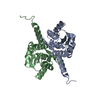
| ||||||||
|---|---|---|---|---|---|---|---|---|---|
| 1 | 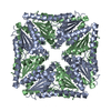
| ||||||||
| 2 | x 8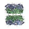
| ||||||||
| Unit cell |
|
-Components
| #1: Protein | Mass: 18858.666 Da / Num. of mol.: 2 / Mutation: H89N Source method: isolated from a genetically manipulated source Source: (gene. exp.) Acetobacter aceti (bacteria) / Strain: 1023 / Gene: purE / Plasmid: pET-23a(+) / Production host: Escherichia coli (E. coli) / Strain (production host): BL21-CodonPlus / References: UniProt: Q2QJL3#2: Water | ChemComp-HOH / | Water Mass: 18.015 Da / Num. of mol.: 276 / Source method: isolated from a natural source / Formula: H2O Mass: 18.015 Da / Num. of mol.: 276 / Source method: isolated from a natural source / Formula: H2O |
|---|
-Experimental details
-Experiment
| Experiment | Method: X-RAY DIFFRACTION / Number of used crystals: 1 |
|---|
- Sample preparation
| Crystal | Density Matthews: 2.68 Å3/Da / Density % sol: 54.04 % |
|---|---|
| Crystal grow | Temperature: 298 K / Method: vapor diffusion, hanging drop / pH: 8 Details: 0.2M lithium sulfate 0.1M Tris pH 8 30% (w/v) PEG 4000 (soaked at pH 7), VAPOR DIFFUSION, HANGING DROP, temperature 298K |
-Data collection
| Diffraction | Mean temperature: 111 K |
|---|---|
| Diffraction source | Source: ROTATING ANODE / Type: RIGAKU RU200 / Wavelength: 1.54 Å |
| Detector | Type: RIGAKU RAXIS IV / Detector: IMAGE PLATE / Date: Oct 5, 2005 |
| Radiation | Monochromator: graphite / Protocol: SINGLE WAVELENGTH / Monochromatic (M) / Laue (L): M / Scattering type: x-ray |
| Radiation wavelength | Wavelength: 1.54 Å / Relative weight: 1 |
| Reflection | Resolution: 1.9→30 Å / Num. obs: 31276 / % possible obs: 95.9 % / Observed criterion σ(I): -3 / Redundancy: 4.9 % / Biso Wilson estimate: 15.6 Å2 / Rsym value: 0.091 / Net I/σ(I): 17.2 |
| Reflection shell | Resolution: 1.9→1.97 Å / Redundancy: 4.6 % / Mean I/σ(I) obs: 3.9 / Num. unique all: 3171 / Rsym value: 0.439 / % possible all: 99 |
- Processing
| Software |
| ||||||||||||||||||||||||||||||||||||||||||||||||||||||||||||||||||||||||||||||||
|---|---|---|---|---|---|---|---|---|---|---|---|---|---|---|---|---|---|---|---|---|---|---|---|---|---|---|---|---|---|---|---|---|---|---|---|---|---|---|---|---|---|---|---|---|---|---|---|---|---|---|---|---|---|---|---|---|---|---|---|---|---|---|---|---|---|---|---|---|---|---|---|---|---|---|---|---|---|---|---|---|---|
| Refinement | Method to determine structure: FOURIER SYNTHESIS Starting model: PDB model 1U11 Resolution: 1.9→29.31 Å / Rfactor Rfree error: 0.005 / Data cutoff high absF: 296467.04 / Data cutoff low absF: 0 / Isotropic thermal model: RESTRAINED / Cross valid method: THROUGHOUT / σ(F): 0 / Stereochemistry target values: Engh & Huber
| ||||||||||||||||||||||||||||||||||||||||||||||||||||||||||||||||||||||||||||||||
| Solvent computation | Solvent model: FLAT MODEL / Bsol: 40.0944 Å2 / ksol: 0.344566 e/Å3 | ||||||||||||||||||||||||||||||||||||||||||||||||||||||||||||||||||||||||||||||||
| Displacement parameters | Biso mean: 19.8 Å2
| ||||||||||||||||||||||||||||||||||||||||||||||||||||||||||||||||||||||||||||||||
| Refine analyze |
| ||||||||||||||||||||||||||||||||||||||||||||||||||||||||||||||||||||||||||||||||
| Refinement step | Cycle: LAST / Resolution: 1.9→29.31 Å
| ||||||||||||||||||||||||||||||||||||||||||||||||||||||||||||||||||||||||||||||||
| Refine LS restraints |
| ||||||||||||||||||||||||||||||||||||||||||||||||||||||||||||||||||||||||||||||||
| LS refinement shell | Resolution: 1.9→2.02 Å / Rfactor Rfree error: 0.017 / Total num. of bins used: 6
| ||||||||||||||||||||||||||||||||||||||||||||||||||||||||||||||||||||||||||||||||
| Xplor file |
|
