 Movie
Movie Controller


+ Open data

- Basic information
| Entry | Database: PDB / ID: 2fpf | ||||||
|---|---|---|---|---|---|---|---|
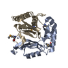
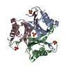
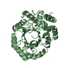
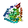
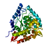
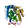
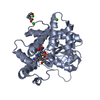
| Title | Crystal structure of the ib1 sh3 dimer at low resolution | ||||||
 Components Components | C-jun-amino-terminal kinase interacting protein 1 | ||||||
 Keywords Keywords |  SIGNALING PROTEIN / SCAFFOLD PROTEIN 1 / ISLET-BRAIN-1 / IB-1 / MITOGEN-ACTIVATED PROTEIN KINASE 8-INTERACTING PROTEIN 1 / JIP-1 RELATED PROTEIN / JRP SIGNALING PROTEIN / SCAFFOLD PROTEIN 1 / ISLET-BRAIN-1 / IB-1 / MITOGEN-ACTIVATED PROTEIN KINASE 8-INTERACTING PROTEIN 1 / JIP-1 RELATED PROTEIN / JRP | ||||||
| Function / homology |  Function and homology information Function and homology informationdentate gyrus mossy fiber / regulation of CD8-positive, alpha-beta T cell proliferation / negative regulation of JUN kinase activity / MAP-kinase scaffold activity / negative regulation of JNK cascade / JUN kinase binding / mitogen-activated protein kinase kinase kinase binding / mitogen-activated protein kinase kinase binding / dendritic growth cone / kinesin binding ...dentate gyrus mossy fiber / regulation of CD8-positive, alpha-beta T cell proliferation / negative regulation of JUN kinase activity / MAP-kinase scaffold activity / negative regulation of JNK cascade / JUN kinase binding / mitogen-activated protein kinase kinase kinase binding / mitogen-activated protein kinase kinase binding / dendritic growth cone / kinesin binding / regulation of JNK cascade / negative regulation of intrinsic apoptotic signaling pathway / axonal growth cone / JNK cascade / vesicle-mediated transport / mitochondrial membrane / positive regulation of JNK cascade / neuron projection / axon / neuronal cell body / dendrite / synapse / endoplasmic reticulum membrane / regulation of DNA-templated transcription / negative regulation of apoptotic process / protein kinase binding / perinuclear region of cytoplasm / signal transduction / membrane / identical protein binding / nucleus / cytosol / cytoplasmSimilarity search - Function | ||||||
| Biological species |  Rattus norvegicus (Norway rat) Rattus norvegicus (Norway rat) | ||||||
| Method | X-RAY DIFFRACTION / SYNCHROTRON / MOLECULAR REPLACEMENT / Resolution: 3 Å | ||||||
 Authors Authors | Kristensen, O. / Dar, I. / Gajhede, M. | ||||||
 Citation Citation | Journal: Embo J. / Year: 2006 Title: A unique set of SH3-SH3 interactions controls IB1 homodimerization Authors: Kristensen, O. / Guenat, S. / Dar, I. / Allaman-Pillet, N. / Abderrahmani, A. / Ferdaoussi, M. / Roduit, R. / Maurer, F. / Beckmann, J.S. / Kastrup, J.S. / Gajhede, M. / Bonny, C. #1: Journal: Science / Year: 1997 Title: A cytoplasmic inhibitor of the JNK signal transduction pathway Authors: Dickens, M. / Rogers, J.S. / Cavanagh, J. / Raitano, A. / Xia, Z. / Halpern, J.R. / Greenberg, M.E. / Sawyers, C.L. / Davis, R.J. #2: Journal: J.Biol.Chem. / Year: 1998 Title: IB1, a JIP-1-related nuclear protein present in insulin-secreting cells Authors: Bonny, C. / Nicod, P. / Waeber, G. #3: Journal: J.Biol.Chem. / Year: 2003 Title: Recruitment of JNK to JIP1 and JNK-dependent JIP1 phosphorylation regulates JNK module dynamics and activation Authors: Nihalani, D. / Wong, H.N. / Holzman, L.B. #4: Journal: Mol.Cell.Biol. / Year: 1999 Title: The JIP group of mitogen-activated protein kinase scaffold proteins Authors: Yasuda, J. / Whitmarsh, A.J. / Cavanagh, J. / Sharma, M. / Davis, R.J. | ||||||
| History |
|
- Structure visualization
| Structure viewer | Molecule: MolmilJmol/JSmol |
|---|
- Downloads & links
-Download
| PDBx/mmCIF format | 2fpf.cif.gz | 57.4 KB | Display | PDBx/mmCIF format |
|---|---|---|---|---|
| PDB format | pdb2fpf.ent.gz | 43.5 KB | Display | PDB format |
| PDBx/mmJSON format | 2fpf.json.gz | Tree view | PDBx/mmJSON format | |
| Others |  Other downloads Other downloads |
-Validation report
| Arichive directory | https://data.pdbj.org/pub/pdb/validation_reports/fp/2fpfftp://data.pdbj.org/pub/pdb/validation_reports/fp/2fpf | HTTPS FTP |
|---|
-Related structure data
| Related structure data | 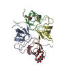 2fpdSC  2fpeC S: Starting model for refinement C: citing same article ( |
|---|---|
| Similar structure data |

-Links
 PDBj
PDBj
- Assembly
| Deposited unit | 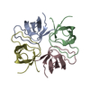
| ||||||||||||||||||||
|---|---|---|---|---|---|---|---|---|---|---|---|---|---|---|---|---|---|---|---|---|---|
| 1 |
| ||||||||||||||||||||
| Unit cell |
| ||||||||||||||||||||
| Noncrystallographic symmetry (NCS) | NCS oper:
|
-Components
| #1: Protein | Mass: 8463.264 Da / Num. of mol.: 4 / Fragment: SH3 DOMAIN, RESIDUES -1-60 Source method: isolated from a genetically manipulated source Source: (gene. exp.) Rattus norvegicus (Norway rat) / Gene: Mapk8ip1, Ib1, Jip1 / Plasmid: PGEX 4T-1 / Production host:  Escherichia coli (E. coli) / Strain (production host): ROSETTA BL21(DE3)PLYSS / References: UniProt: Q9R237 Escherichia coli (E. coli) / Strain (production host): ROSETTA BL21(DE3)PLYSS / References: UniProt: Q9R237 |
|---|
-Experimental details
-Experiment
| Experiment | Method: X-RAY DIFFRACTION / Number of used crystals: 1 |
|---|
- Sample preparation
| Crystal | Density Matthews: 2.8 Å3/Da / Density % sol: 56 % |
|---|---|
| Crystal grow | Temperature: 293 K / Method: vapor diffusion, hanging drop / pH: 6.5 Details: SODIUM CITRATE, HEPES, HYDROGEN PEROXIDE, pH 6.50, VAPOR DIFFUSION, HANGING DROP, temperature 293K |
-Data collection
| Diffraction | Mean temperature: 100 K |
|---|---|
| Diffraction source | Source: SYNCHROTRON / Site: ESRF  / Beamline: ID29 / Wavelength: 0.979 / Beamline: ID29 / Wavelength: 0.979 |
| Detector | Type: ADSC QUANTUM 210 / Detector: CCD / Date: Feb 22, 2003 |
| Radiation | Protocol: SINGLE WAVELENGTH / Monochromatic (M) / Laue (L): M / Scattering type: x-ray |
| Radiation wavelength | Wavelength: 0.979 Å / Relative weight: 1 |
| Reflection | Resolution: 3→30 Å / Num. obs: 8075 / % possible obs: 99.2 % / Observed criterion σ(I): -3 / Redundancy: 4.6 % / Biso Wilson estimate: 48 Å2 / Rmerge(I) obs: 0.102 / Rsym value: 0.102 / Net I/σ(I): 12.07 |
| Reflection shell | Resolution: 3→3.11 Å / Redundancy: 4.5 % / Rmerge(I) obs: 0.461 / Mean I/σ(I) obs: 2.9 / Rsym value: 0.461 / % possible all: 100 |
- Processing
| Software |
| ||||||||||||||||||||||||||||||||||||||||||||||||||||||||||||||||||||||||||||||||
|---|---|---|---|---|---|---|---|---|---|---|---|---|---|---|---|---|---|---|---|---|---|---|---|---|---|---|---|---|---|---|---|---|---|---|---|---|---|---|---|---|---|---|---|---|---|---|---|---|---|---|---|---|---|---|---|---|---|---|---|---|---|---|---|---|---|---|---|---|---|---|---|---|---|---|---|---|---|---|---|---|---|
| Refinement | Method to determine structure: MOLECULAR REPLACEMENT Starting model: PDB ENTRY 2FPD Resolution: 3→28.77 Å / Rfactor Rfree error: 0.01 / Data cutoff high absF: 230208.16 / Data cutoff low absF: 0 / Isotropic thermal model: RESTRAINED / Cross valid method: THROUGHOUT / σ(F): 0 / Stereochemistry target values: Engh & Huber
| ||||||||||||||||||||||||||||||||||||||||||||||||||||||||||||||||||||||||||||||||
| Solvent computation | Solvent model: FLAT MODEL / Bsol: 39.58 Å2 / ksol: 0.4 e/Å3 | ||||||||||||||||||||||||||||||||||||||||||||||||||||||||||||||||||||||||||||||||
| Displacement parameters | Biso mean: 51 Å2
| ||||||||||||||||||||||||||||||||||||||||||||||||||||||||||||||||||||||||||||||||
| Refine analyze |
| ||||||||||||||||||||||||||||||||||||||||||||||||||||||||||||||||||||||||||||||||
| Refinement step | Cycle: LAST / Resolution: 3→28.77 Å
| ||||||||||||||||||||||||||||||||||||||||||||||||||||||||||||||||||||||||||||||||
| Refine LS restraints |
| ||||||||||||||||||||||||||||||||||||||||||||||||||||||||||||||||||||||||||||||||
| Refine LS restraints NCS | NCS model details: CONSTR | ||||||||||||||||||||||||||||||||||||||||||||||||||||||||||||||||||||||||||||||||
| LS refinement shell | Resolution: 3→3.19 Å / Rfactor Rfree error: 0.034 / Total num. of bins used: 6
| ||||||||||||||||||||||||||||||||||||||||||||||||||||||||||||||||||||||||||||||||
| Xplor file | Serial no: 1 / Param file: PROTEIN_REP.PARAM / Topol file: PROTEIN.TOP |
