 Movie
Movie Controller

[English] 日本語
 Yorodumi
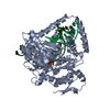
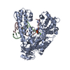
- PDB-2flf: Crystal structure of l-fuculose-1-phosphate aldolase from Thermus... -
+ Open data

- Basic information
| Entry | Database: PDB / ID: 2flf | ||||||
|---|---|---|---|---|---|---|---|
| Title | Crystal structure of l-fuculose-1-phosphate aldolase from Thermus Thermophilus HB8 | ||||||
 Components Components | fuculose-1-phosphate aldolase | ||||||
 Keywords Keywords |  LYASE / Class II Aldolase / metal binding / fuculose phosphate / RIKEN STRUCTURAL GENOMICS/PROTEOMICS INITIATIVE / RSGI / NPPSFA / NATIONAL PROJECT ON PROTEIN STRUCTURAL AND FUNCTIONAL ANALYSES LYASE / Class II Aldolase / metal binding / fuculose phosphate / RIKEN STRUCTURAL GENOMICS/PROTEOMICS INITIATIVE / RSGI / NPPSFA / NATIONAL PROJECT ON PROTEIN STRUCTURAL AND FUNCTIONAL ANALYSES | ||||||
| Function / homology |  Function and homology information Function and homology information | ||||||
| Biological species |   Thermus thermophilus (bacteria) Thermus thermophilus (bacteria) | ||||||
| Method | X-RAY DIFFRACTION / SYNCHROTRON / MOLECULAR REPLACEMENT / Resolution: 2.7 Å | ||||||
 Authors Authors | Jeyakanthan, J. / Yokoyama, S. / Shiro, Y. / RIKEN Structural Genomics/Proteomics Initiative (RSGI) | ||||||
 Citation Citation | Journal: Acta Crystallogr.,Sect.F / Year: 2005 Title: Purification, crystallization and preliminary X-ray crystallographic study of the L-fuculose-1-phosphate aldolase (FucA) from Thermus thermophilus HB8 Authors: Jeyakanthan, J. / Taka, J. / Kikuchi, A. / Kuroishi, C. / Yutani, K. / Shiro, Y. | ||||||
| History |
|
- Structure visualization
| Structure viewer | Molecule: MolmilJmol/JSmol |
|---|
- Downloads & links
-Download
| PDBx/mmCIF format | 2flf.cif.gz | 297.3 KB | Display | PDBx/mmCIF format |
|---|---|---|---|---|
| PDB format | pdb2flf.ent.gz | 243.2 KB | Display | PDB format |
| PDBx/mmJSON format | 2flf.json.gz | Tree view | PDBx/mmJSON format | |
| Others |  Other downloads Other downloads |
-Validation report
| Arichive directory | https://data.pdbj.org/pub/pdb/validation_reports/fl/2flfftp://data.pdbj.org/pub/pdb/validation_reports/fl/2flf | HTTPS FTP |
|---|
-Related structure data
| Related structure data |  2fk5SC S: Starting model for refinement C: citing same article ( |
|---|---|
| Similar structure data | |
| Other databases |


-Links
 PDBj
PDBj- Assembly
| Deposited unit | 
| ||||||||
|---|---|---|---|---|---|---|---|---|---|
| 1 | 
| ||||||||
| 2 | 
| ||||||||
| Unit cell |
|
-Components
| #1: Protein | Mass: 21620.785 Da / Num. of mol.: 8 Source method: isolated from a genetically manipulated source Source: (gene. exp.) Thermus thermophilus (bacteria) / Strain: HB8 / Plasmid: PET-11A / Production host: Escherichia coli (E. coli) / References: UniProt: Q5SHB9, L-fuculose-phosphate aldolase#2: Water | ChemComp-HOH / | Water Mass: 18.015 Da / Num. of mol.: 257 / Source method: isolated from a natural source / Formula: H2O Mass: 18.015 Da / Num. of mol.: 257 / Source method: isolated from a natural source / Formula: H2O |
|---|
-Experimental details
-Experiment
| Experiment | Method: X-RAY DIFFRACTION / Number of used crystals: 1 |
|---|
- Sample preparation
| Crystal | Density Matthews: 2.17 Å3/Da / Density % sol: 43.27 % |
|---|---|
| Crystal grow | Temperature: 295 K / Method: vapor diffusion, sitting drop / pH: 6 Details: 28% PEG 4000, 0.1M CITRATE-NaOH, pH 6.0, VAPOR DIFFUSION, SITTING DROP, temperature 295K |
-Data collection
| Diffraction | Mean temperature: 100 K |
|---|---|
| Diffraction source | Source: SYNCHROTRON / Site: SPring-8  / Beamline: BL26B1 / Wavelength: 1 Å / Beamline: BL26B1 / Wavelength: 1 Å |
| Detector | Type: RIGAKU RAXIS IV / Detector: IMAGE PLATE / Date: Jun 25, 2003 / Details: mirrors |
| Radiation | Monochromator: GRAPHITE / Protocol: SINGLE WAVELENGTH / Monochromatic (M) / Laue (L): M / Scattering type: x-ray |
| Radiation wavelength | Wavelength: 1 Å / Relative weight: 1 |
| Reflection | Resolution: 2.6→30 Å / Num. obs: 46644 / % possible obs: 99.5 % / Observed criterion σ(F): 0 / Observed criterion σ(I): 0 / Biso Wilson estimate: 45.4 Å2 / Rmerge(I) obs: 0.074 |
| Reflection shell | Resolution: 2.6→2.69 Å / Rmerge(I) obs: 0.564 / % possible all: 99.9 |
- Processing
| Software |
| ||||||||||||||||||||||||||||||||||||||||||||||||||||||||||||||||||||||||||||||||
|---|---|---|---|---|---|---|---|---|---|---|---|---|---|---|---|---|---|---|---|---|---|---|---|---|---|---|---|---|---|---|---|---|---|---|---|---|---|---|---|---|---|---|---|---|---|---|---|---|---|---|---|---|---|---|---|---|---|---|---|---|---|---|---|---|---|---|---|---|---|---|---|---|---|---|---|---|---|---|---|---|---|
| Refinement | Method to determine structure: MOLECULAR REPLACEMENT Starting model: PDB ENTRY 2FK5 Resolution: 2.7→19.97 Å / Rfactor Rfree error: 0.007 / Data cutoff high absF: 512801.78 / Data cutoff low absF: 0 / Isotropic thermal model: RESTRAINED / Cross valid method: THROUGHOUT / σ(F): 0 / Stereochemistry target values: Engh & Huber
| ||||||||||||||||||||||||||||||||||||||||||||||||||||||||||||||||||||||||||||||||
| Solvent computation | Solvent model: FLAT MODEL / Bsol: 28.1263 Å2 / ksol: 0.260732 e/Å3 | ||||||||||||||||||||||||||||||||||||||||||||||||||||||||||||||||||||||||||||||||
| Displacement parameters | Biso mean: 61.5 Å2
| ||||||||||||||||||||||||||||||||||||||||||||||||||||||||||||||||||||||||||||||||
| Refine analyze |
| ||||||||||||||||||||||||||||||||||||||||||||||||||||||||||||||||||||||||||||||||
| Refinement step | Cycle: LAST / Resolution: 2.7→19.97 Å
| ||||||||||||||||||||||||||||||||||||||||||||||||||||||||||||||||||||||||||||||||
| Refine LS restraints |
| ||||||||||||||||||||||||||||||||||||||||||||||||||||||||||||||||||||||||||||||||
| LS refinement shell | Resolution: 2.7→2.87 Å / Rfactor Rfree error: 0.025 / Total num. of bins used: 6
| ||||||||||||||||||||||||||||||||||||||||||||||||||||||||||||||||||||||||||||||||
| Xplor file |
|
