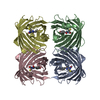
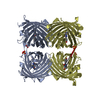
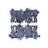
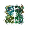
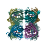
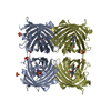
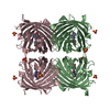
- PDB-2fl1: Crystal structure of red fluorescent protein from Zoanthus, zRFP5... -
+
Open data
ID or keywords:
Loading...
-
Basic information
Entry
Database: PDB / ID: 2fl1
Title
Crystal structure of red fluorescent protein from Zoanthus, zRFP574, at 2.4A resolution
Components
Red fluorescent protein zoanRFP
Keywords
FLUORESCENT PROTEIN / Red fluorescent protein / button polyp / Zoanthus sp. / chromophore / beta-can fold / beta barrel / tightly packed tetramer / intersubunit interface / fluorescent marker / emission maximum 574nm / zRFP574
Function / homology
Green Fluorescent Protein / Green fluorescent protein / Green fluorescent protein-related / Green fluorescent protein / Green fluorescent protein / bioluminescence / Beta Barrel / Mainly Beta / Red fluorescent protein zoanRFP
Function and homology information
Biological species
Zoanthus sp. (invertebrata)
Method
X-RAY DIFFRACTION / MOLECULAR REPLACEMENT / Resolution: 2.4 Å
SEQUENCE Chromophore (XYG 66) is formed autocatalytically from Asp-Tyr-Gly (according to nucleotide ...SEQUENCE Chromophore (XYG 66) is formed autocatalytically from Asp-Tyr-Gly (according to nucleotide sequence) after decarboxylation of Asp and other deprotonation processes.
A: Red fluorescent protein zoanRFP B: Red fluorescent protein zoanRFP C: Red fluorescent protein zoanRFP D: Red fluorescent protein zoanRFP hetero molecules
In the structure databanks used in Yorodumi, some data are registered as the other names, "COVID-19 virus" and "2019-nCoV". Here are the details of the virus and the list of structure data.
Jan 31, 2019. EMDB accession codes are about to change! (news from PDBe EMDB page)
EMDB accession codes are about to change! (news from PDBe EMDB page)
The allocation of 4 digits for EMDB accession codes will soon come to an end. Whilst these codes will remain in use, new EMDB accession codes will include an additional digit and will expand incrementally as the available range of codes is exhausted. The current 4-digit format prefixed with “EMD-” (i.e. EMD-XXXX) will advance to a 5-digit format (i.e. EMD-XXXXX), and so on. It is currently estimated that the 4-digit codes will be depleted around Spring 2019, at which point the 5-digit format will come into force.
The EM Navigator/Yorodumi systems omit the EMD- prefix.
Related info.:Q: What is EMD? / ID/Accession-code notation in Yorodumi/EM Navigator
Yorodumi is a browser for structure data from EMDB, PDB, SASBDB, etc.
This page is also the successor to EM Navigator detail page, and also detail information page/front-end page for Omokage search.
The word "yorodu" (or yorozu) is an old Japanese word meaning "ten thousand". "mi" (miru) is to see.
Related info.:EMDB / PDB / SASBDB / Comparison of 3 databanks / Yorodumi Search / Aug 31, 2016. New EM Navigator & Yorodumi / Yorodumi Papers / Jmol/JSmol / Function and homology information / Changes in new EM Navigator and Yorodumi
 Movie
Movie Controller

 Yorodumi
 Open data
 Basic information
 Components
 Keywords
 FLUORESCENT PROTEIN /
  Function and homology information
 Zoanthus sp. (invertebrata)
 Authors
 Citation
 Structure visualization
 Downloads & links
 Other downloads


 PDBj
PDBj

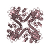
 Assembly


 Mass: 96.063 Da / Num. of mol.: 9 / Source method: obtained synthetically / Formula: SO4
 Mass: 18.015 Da / Num. of mol.: 412 / Source method: isolated from a natural source / Formula: H2O
 Sample preparation
 Processing