 Movie
Movie Controller


+ Open data

- Basic information
| Entry | Database: PDB / ID: 2fau | ||||||
|---|---|---|---|---|---|---|---|
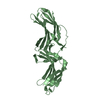
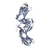
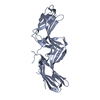
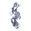
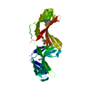
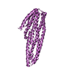
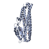
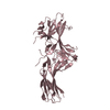
| Title | Crystal structure of human vps26 | ||||||
 Components Components | Vacuolar protein sorting 26 Vacuole Vacuole | ||||||
 Keywords Keywords | PROTEIN TRANSPORT / ARRESTIN / RETROMER | ||||||
| Function / homology |  Function and homology information Function and homology informationtubular endosome / retromer, cargo-selective complex / WNT ligand biogenesis and trafficking / retromer complex / endocytic recycling / retrograde transport, endosome to Golgi / regulation of macroautophagy / intracellular protein transport / vesicle / lysosome ...tubular endosome / retromer, cargo-selective complex / WNT ligand biogenesis and trafficking / retromer complex / endocytic recycling / retrograde transport, endosome to Golgi / regulation of macroautophagy / intracellular protein transport / vesicle / lysosome / early endosome / endosome membrane / endosome / cytosolSimilarity search - Function | ||||||
| Biological species |  Homo sapiens (human) Homo sapiens (human) | ||||||
| Method | X-RAY DIFFRACTION / SYNCHROTRON / AB INITIO PHASING / Resolution: 2.1 Å | ||||||
 Authors Authors | Shi, H. / Rojas, R. / Bonifacino, J.S. / Hurley, J.H. | ||||||
 Citation Citation | Journal: Nat.Struct.Mol.Biol. / Year: 2006 Title: The retromer subunit Vps26 has an arrestin fold and binds Vps35 through its C-terminal domain. Authors: Shi, H. / Rojas, R. / Bonifacino, J.S. / Hurley, J.H. | ||||||
| History |
|
- Structure visualization
| Structure viewer | Molecule: MolmilJmol/JSmol |
|---|
- Downloads & links
-Download
| PDBx/mmCIF format | 2fau.cif.gz | 71.3 KB | Display | PDBx/mmCIF format |
|---|---|---|---|---|
| PDB format | pdb2fau.ent.gz | 56.3 KB | Display | PDB format |
| PDBx/mmJSON format | 2fau.json.gz | Tree view | PDBx/mmJSON format | |
| Others |  Other downloads Other downloads |
-Validation report
| Arichive directory | https://data.pdbj.org/pub/pdb/validation_reports/fa/2fauftp://data.pdbj.org/pub/pdb/validation_reports/fa/2fau | HTTPS FTP |
|---|
-Related structure data
| Similar structure data |
|---|


-Links
 PDBj
PDBj- Assembly
| Deposited unit | 
| ||||||||
|---|---|---|---|---|---|---|---|---|---|
| 1 |
| ||||||||
| Unit cell |
|
-Components
| #1: Protein | Vacuole / Vesicle protein sorting 26 / hVPS26 Mass: 40010.609 Da / Num. of mol.: 1 Source method: isolated from a genetically manipulated source Source: (gene. exp.) Homo sapiens (human) / Gene: VPS26A, VPS26 / Plasmid: pmr101 / Production host:  Escherichia coli (E. coli) / Strain (production host): BL21 (DE3) Gold / References: UniProt: O75436 Escherichia coli (E. coli) / Strain (production host): BL21 (DE3) Gold / References: UniProt: O75436 |
|---|---|
| #2: Chemical | ChemComp-GOL / Glycerol  Mass: 92.094 Da / Num. of mol.: 1 / Source method: obtained synthetically / Formula: C3H8O3 Mass: 92.094 Da / Num. of mol.: 1 / Source method: obtained synthetically / Formula: C3H8O3 |
| #3: Water | ChemComp-HOH / Water Mass: 18.015 Da / Num. of mol.: 82 / Source method: isolated from a natural source / Formula: H2O Mass: 18.015 Da / Num. of mol.: 82 / Source method: isolated from a natural source / Formula: H2O |
-Experimental details
-Experiment
| Experiment | Method: X-RAY DIFFRACTION / Number of used crystals: 1 |
|---|
- Sample preparation
| Crystal | Density Matthews: 2.47 Å3/Da / Density % sol: 50.24 % |
|---|---|
| Crystal grow | Temperature: 300 K / Method: vapor diffusion, sitting drop / pH: 6.5 Details: 8% PEG 8000 100mM Na cacodylate pH 6.5 15% glycerol 10mM DTT 1mM TmCl3, VAPOR DIFFUSION, SITTING DROP, temperature 300K |
-Data collection
| Diffraction |
| ||||||||||||||||||||||||
|---|---|---|---|---|---|---|---|---|---|---|---|---|---|---|---|---|---|---|---|---|---|---|---|---|---|
| Diffraction source | Source: SYNCHROTRON / Site: APS  / Beamline: 22-ID / Wavelength: 1.072 Å / Beamline: 22-ID / Wavelength: 1.072 Å | ||||||||||||||||||||||||
| Detector | Type: MARRESEARCH / Detector: CCD / Date: Feb 6, 2005 | ||||||||||||||||||||||||
| Radiation |
| ||||||||||||||||||||||||
| Radiation wavelength | Wavelength: 1.072 Å / Relative weight: 1 | ||||||||||||||||||||||||
| Reflection | Resolution: 2.09→48.71 Å / Num. all: 22533 / Num. obs: 22533 / % possible obs: 87.9 % / Observed criterion σ(F): -3 / Observed criterion σ(I): -3 / Biso Wilson estimate: 21 Å2 | ||||||||||||||||||||||||
| Reflection shell | Resolution: 2.09→2.16 Å / % possible all: 55.4 |
- Processing
| Software |
| ||||||||||||||||||||||||||||||||||||||||||||||||||||||||||||
|---|---|---|---|---|---|---|---|---|---|---|---|---|---|---|---|---|---|---|---|---|---|---|---|---|---|---|---|---|---|---|---|---|---|---|---|---|---|---|---|---|---|---|---|---|---|---|---|---|---|---|---|---|---|---|---|---|---|---|---|---|---|
| Refinement | Method to determine structure: AB INITIO PHASING / Resolution: 2.1→48.71 Å / Rfactor Rfree error: 0.006 / Data cutoff high absF: 860282.46 / Data cutoff low absF: 0 / Isotropic thermal model: RESTRAINED / Cross valid method: THROUGHOUT / σ(F): 0 / Stereochemistry target values: Engh & Huber
| ||||||||||||||||||||||||||||||||||||||||||||||||||||||||||||
| Solvent computation | Solvent model: FLAT MODEL / Bsol: 61.0291 Å2 / ksol: 0.342588 e/Å3 | ||||||||||||||||||||||||||||||||||||||||||||||||||||||||||||
| Displacement parameters | Biso mean: 66.4 Å2
| ||||||||||||||||||||||||||||||||||||||||||||||||||||||||||||
| Refine analyze |
| ||||||||||||||||||||||||||||||||||||||||||||||||||||||||||||
| Refinement step | Cycle: LAST / Resolution: 2.1→48.71 Å
| ||||||||||||||||||||||||||||||||||||||||||||||||||||||||||||
| Refine LS restraints |
| ||||||||||||||||||||||||||||||||||||||||||||||||||||||||||||
| LS refinement shell | Resolution: 2.1→2.23 Å / Rfactor Rfree error: 0.025 / Total num. of bins used: 6
| ||||||||||||||||||||||||||||||||||||||||||||||||||||||||||||
| Xplor file |
|
