 Movie
Movie Controller

[English] 日本語
 Yorodumi
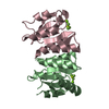
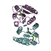
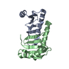
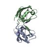
- PDB-2f9h: The Crystal Structure of PTS System IIA Component from Enterococc... -
+ Open data

- Basic information
| Entry | Database: PDB / ID: 2f9h | ||||||
|---|---|---|---|---|---|---|---|
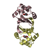
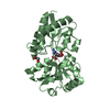
| Title | The Crystal Structure of PTS System IIA Component from Enterococcus faecalis V583 | ||||||
 Components Components | PTS system, IIA component | ||||||
 Keywords Keywords |  STRUCTURAL GENOMICS / UNKNOWN FUNCTION / alpha-beta structure / beta-barrel / dimer / PSI / Protein Structure Initiative / Midwest Center for Structural Genomics / MCSG STRUCTURAL GENOMICS / UNKNOWN FUNCTION / alpha-beta structure / beta-barrel / dimer / PSI / Protein Structure Initiative / Midwest Center for Structural Genomics / MCSG | ||||||
| Function / homology |  Function and homology information Function and homology informationprotein-N(PI)-phosphohistidine-sugar phosphotransferase activity / phosphoenolpyruvate-dependent sugar phosphotransferase system / cytoplasmSimilarity search - Function | ||||||
| Biological species |   Enterococcus faecalis (bacteria) Enterococcus faecalis (bacteria) | ||||||
| Method | X-RAY DIFFRACTION / SYNCHROTRON / MAD / Resolution: 1.57 Å | ||||||
 Authors Authors | Kim, Y. / Quartey, P. / Moy, S. / Bargassa, M. / Collart, F. / Joachimiak, A. / Midwest Center for Structural Genomics (MCSG) | ||||||
 Citation Citation | Journal: To be Published / Year: 2006 Title: The Crystal Structure of PTS System IIA Component from Enterococcus faecalis V583 Authors: Kim, Y. / Quartey, P. / Abdullah, J. / Collart, F. / Joachimiak, A. | ||||||
| History |
|
- Structure visualization
| Structure viewer | Molecule: MolmilJmol/JSmol |
|---|
- Downloads & links
-Download
| PDBx/mmCIF format | 2f9h.cif.gz | 60.2 KB | Display | PDBx/mmCIF format |
|---|---|---|---|---|
| PDB format | pdb2f9h.ent.gz | 48 KB | Display | PDB format |
| PDBx/mmJSON format | 2f9h.json.gz | Tree view | PDBx/mmJSON format | |
| Others |  Other downloads Other downloads |
-Validation report
| Arichive directory | https://data.pdbj.org/pub/pdb/validation_reports/f9/2f9hftp://data.pdbj.org/pub/pdb/validation_reports/f9/2f9h | HTTPS FTP |
|---|
-Related structure data
| Similar structure data | |
|---|---|
| Other databases |


-Links
 PDBj
PDBj- Assembly
| Deposited unit | 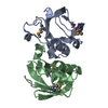
| ||||||||
|---|---|---|---|---|---|---|---|---|---|
| 1 |
| ||||||||
| Unit cell |
|
-Components
| #1: Protein | Mass: 14185.389 Da / Num. of mol.: 2 Source method: isolated from a genetically manipulated source Source: (gene. exp.) Enterococcus faecalis (bacteria) / Strain: V583 / Plasmid: pMCSG7 / Species (production host): Escherichia coli / Production host: Escherichia coli BL21(DE3) (bacteria) / Strain (production host): BL21DE3 / References: GenBank: 29377088, UniProt: Q831B0*PLUS#2: Water | ChemComp-HOH / | Water Mass: 18.015 Da / Num. of mol.: 257 / Source method: isolated from a natural source / Formula: H2O Mass: 18.015 Da / Num. of mol.: 257 / Source method: isolated from a natural source / Formula: H2O |
|---|
-Experimental details
-Experiment
| Experiment | Method: X-RAY DIFFRACTION / Number of used crystals: 2 |
|---|
- Sample preparation
| Crystal | Density Matthews: 2.42 Å3/Da / Density % sol: 49.25 % |
|---|---|
| Crystal grow | Temperature: 291 K / Method: vapor diffusion, sitting drop / pH: 4.6 Details: 0.1 M Sodium acetate pH 4.6, 4% PEG4000, VAPOR DIFFUSION, SITTING DROP, temperature 291K |
-Data collection
| Diffraction |
| ||||||||||||||||||
|---|---|---|---|---|---|---|---|---|---|---|---|---|---|---|---|---|---|---|---|
| Diffraction source |
| ||||||||||||||||||
| Detector |
| ||||||||||||||||||
| Radiation |
| ||||||||||||||||||
| Radiation wavelength |
| ||||||||||||||||||
| Reflection | Resolution: 1.57→34.32 Å / Num. all: 39333 / Num. obs: 38988 / % possible obs: 98.7 % / Observed criterion σ(F): 0 / Observed criterion σ(I): 0 / Redundancy: 12.7 % / Biso Wilson estimate: 23.2 Å2 / Rmerge(I) obs: 0.054 / Net I/σ(I): 17.3 | ||||||||||||||||||
| Reflection shell | Resolution: 1.57→1.63 Å / Redundancy: 8.2 % / Rmerge(I) obs: 0.332 / Mean I/σ(I) obs: 4.5 / Num. unique all: 3411 / % possible all: 88.4 |

- Processing
| Software |
| ||||||||||||||||||||||||||||||||||||||||||||||||||||||||||||||||||||||||||||||||
|---|---|---|---|---|---|---|---|---|---|---|---|---|---|---|---|---|---|---|---|---|---|---|---|---|---|---|---|---|---|---|---|---|---|---|---|---|---|---|---|---|---|---|---|---|---|---|---|---|---|---|---|---|---|---|---|---|---|---|---|---|---|---|---|---|---|---|---|---|---|---|---|---|---|---|---|---|---|---|---|---|---|
| Refinement | Method to determine structure: MAD / Resolution: 1.57→34.32 Å / Rfactor Rfree error: 0.005 / Data cutoff high absF: 1411320.03 / Data cutoff low absF: 0 / Isotropic thermal model: RESTRAINED / Cross valid method: THROUGHOUT / σ(F): 0 / Stereochemistry target values: Engh & Huber Details: Crystal was twinned; fraction 0.442, direction -h,-k,l
| ||||||||||||||||||||||||||||||||||||||||||||||||||||||||||||||||||||||||||||||||
| Solvent computation | Solvent model: FLAT MODEL / Bsol: 45.0654 Å2 / ksol: 0.348908 e/Å3 | ||||||||||||||||||||||||||||||||||||||||||||||||||||||||||||||||||||||||||||||||
| Displacement parameters | Biso mean: 25.4 Å2
| ||||||||||||||||||||||||||||||||||||||||||||||||||||||||||||||||||||||||||||||||
| Refine analyze |
| ||||||||||||||||||||||||||||||||||||||||||||||||||||||||||||||||||||||||||||||||
| Refinement step | Cycle: LAST / Resolution: 1.57→34.32 Å
| ||||||||||||||||||||||||||||||||||||||||||||||||||||||||||||||||||||||||||||||||
| Refine LS restraints |
| ||||||||||||||||||||||||||||||||||||||||||||||||||||||||||||||||||||||||||||||||
| LS refinement shell | Resolution: 1.57→1.64 Å / Rfactor Rfree error: 0.016 / Total num. of bins used: 8
| ||||||||||||||||||||||||||||||||||||||||||||||||||||||||||||||||||||||||||||||||
| Xplor file |
|
