 Movie
Movie Controller

[English] 日本語
 Yorodumi
- PDB-2f60: Crystal Structure of the Dihydrolipoamide Dehydrogenase (E3)-Bind... -
+ Open data

- Basic information
| Entry | Database: PDB / ID: 2f60 | ||||||
|---|---|---|---|---|---|---|---|
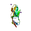
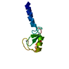
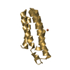
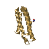
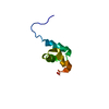
| Title | Crystal Structure of the Dihydrolipoamide Dehydrogenase (E3)-Binding Domain of Human E3-Binding Protein | ||||||
 Components Components | Pyruvate dehydrogenase protein X component | ||||||
 Keywords Keywords | PROTEIN BINDING / protein-binding protein / E3BD | ||||||
| Function / homology |  Function and homology information Function and homology informationacetyl-CoA biosynthetic process from pyruvate / pyruvate dehydrogenase complex / : / Pyruvate metabolism / Glyoxylate metabolism and glycine degradation / Regulation of pyruvate dehydrogenase (PDH) complex / Signaling by Retinoic Acid / acyltransferase activity / mitochondrial matrix / mitochondrionSimilarity search - Function | ||||||
| Biological species |  Homo sapiens (human) Homo sapiens (human) | ||||||
| Method | X-RAY DIFFRACTION / SYNCHROTRON / MOLECULAR REPLACEMENT / Resolution: 1.55 Å | ||||||
 Authors Authors | Brautigam, C.A. / Chuang, J.L. / Wynn, R.M. / Tomchick, D.R. / Machius, M. / Chuang, D.T. | ||||||
 Citation Citation | Journal: Structure / Year: 2006 Title: Structural Insight into Interactions between Dihydrolipoamide Dehydrogenase (E3) and E3 Binding Protein of Human Pyruvate Dehydrogenase Complex. Authors: Brautigam, C.A. / Wynn, R.M. / Chuang, J.L. / Machius, M. / Tomchick, D.R. / Chuang, D.T. | ||||||
| History |
|
- Structure visualization
| Structure viewer | Molecule: MolmilJmol/JSmol |
|---|
- Downloads & links
-Download
| PDBx/mmCIF format | 2f60.cif.gz | 26.3 KB | Display | PDBx/mmCIF format |
|---|---|---|---|---|
| PDB format | pdb2f60.ent.gz | 16.9 KB | Display | PDB format |
| PDBx/mmJSON format | 2f60.json.gz | Tree view | PDBx/mmJSON format | |
| Others |  Other downloads Other downloads |
-Validation report
| Arichive directory | https://data.pdbj.org/pub/pdb/validation_reports/f6/2f60ftp://data.pdbj.org/pub/pdb/validation_reports/f6/2f60 | HTTPS FTP |
|---|
-Related structure data
| Related structure data |  2f5zSC S: Starting model for refinement C: citing same article ( |
|---|---|
| Similar structure data |


-Links
 PDBj
PDBj


- Assembly
| Deposited unit | 
| ||||||||
|---|---|---|---|---|---|---|---|---|---|
| 1 |
| ||||||||
| Unit cell |
|
-Components
| #1: Protein | / Dihydrolipoamide dehydrogenase-binding protein of pyruvate dehydrogenase complex / Lipoyl- ...Dihydrolipoamide dehydrogenase-binding protein of pyruvate dehydrogenase complex / Lipoyl-containing pyruvate dehydrogenase complex component X / E3-binding protein / E3BP / proX Mass: 7150.151 Da / Num. of mol.: 1 / Fragment: E3-binding domain, residues 173-230 / Mutation: K120G, T176L Source method: isolated from a genetically manipulated source Source: (gene. exp.) Homo sapiens (human) / Gene: PDHX, PDX1 / Plasmid: pET-LTE3BD / Species (production host): Escherichia coli / Production host:  Escherichia coli BL21 (bacteria) / Strain (production host): BL21 / References: UniProt: O00330 Escherichia coli BL21 (bacteria) / Strain (production host): BL21 / References: UniProt: O00330 | ||
|---|---|---|---|
| #2: Chemical | Glycerol  Mass: 92.094 Da / Num. of mol.: 2 / Source method: obtained synthetically / Formula: C3H8O3 Mass: 92.094 Da / Num. of mol.: 2 / Source method: obtained synthetically / Formula: C3H8O3#3: Water | ChemComp-HOH / | Water Mass: 18.015 Da / Num. of mol.: 63 / Source method: isolated from a natural source / Formula: H2O Mass: 18.015 Da / Num. of mol.: 63 / Source method: isolated from a natural source / Formula: H2O |
-Experimental details
-Experiment
| Experiment | Method: X-RAY DIFFRACTION / Number of used crystals: 1 |
|---|
- Sample preparation
| Crystal | Density Matthews: 2.62 Å3/Da / Density % sol: 53.11 % |
|---|---|
| Crystal grow | Temperature: 293 K / pH: 6.3 Details: 1.35 M sodium citrate, 0.075% beta-octyl-glucopyranoside, pH 6.3, VAPOR DIFFUSION, HANGING DROP, temperature 293K, pH 6.30 |
-Data collection
| Diffraction | Mean temperature: 100 K |
|---|---|
| Diffraction source | Source: SYNCHROTRON / Site: APS  / Beamline: 19-ID / Wavelength: 0.9793 / Beamline: 19-ID / Wavelength: 0.9793 |
| Detector | Type: ADSC QUANTUM 315 / Detector: CCD / Date: Nov 12, 2004 |
| Radiation | Monochromator: SI (111) / Protocol: SINGLE WAVELENGTH / Monochromatic (M) / Laue (L): M / Scattering type: x-ray |
| Radiation wavelength | Wavelength: 0.9793 Å / Relative weight: 1 |
| Reflection | Resolution: 1.55→28.5 Å / Num. obs: 12109 / % possible obs: 98.9 % / Observed criterion σ(I): -3 / Redundancy: 13.7 % / Biso Wilson estimate: 23.1 Å2 / Rsym value: 0.032 / Net I/σ(I): 67.8 |
| Reflection shell | Resolution: 1.55→1.61 Å / Redundancy: 7.9 % / Mean I/σ(I) obs: 8.6 / Rsym value: 0.148 / % possible all: 95.4 |
- Processing
| Software |
| ||||||||||||||||||||||||||||||||||||||||||||||||||||||||||||
|---|---|---|---|---|---|---|---|---|---|---|---|---|---|---|---|---|---|---|---|---|---|---|---|---|---|---|---|---|---|---|---|---|---|---|---|---|---|---|---|---|---|---|---|---|---|---|---|---|---|---|---|---|---|---|---|---|---|---|---|---|---|
| Refinement | Method to determine structure: MOLECULAR REPLACEMENT Starting model: PDB ENTRY 2F5Z Resolution: 1.55→28.5 Å / Isotropic thermal model: RESTRAINED / Cross valid method: THROUGHOUT / σ(F): 0 / Stereochemistry target values: ENGH & HUBER
| ||||||||||||||||||||||||||||||||||||||||||||||||||||||||||||
| Displacement parameters | Biso mean: 26 Å2
| ||||||||||||||||||||||||||||||||||||||||||||||||||||||||||||
| Refine analyze |
| ||||||||||||||||||||||||||||||||||||||||||||||||||||||||||||
| Refinement step | Cycle: LAST / Resolution: 1.55→28.5 Å
| ||||||||||||||||||||||||||||||||||||||||||||||||||||||||||||
| Refine LS restraints |
| ||||||||||||||||||||||||||||||||||||||||||||||||||||||||||||
| LS refinement shell | Resolution: 1.55→1.65 Å / Rfactor Rfree error: 0.026
|
