 Movie
Movie Controller


+ Open data

- Basic information
| Entry | Database: PDB / ID: 2f5y | ||||||
|---|---|---|---|---|---|---|---|
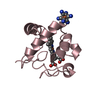
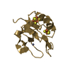
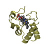
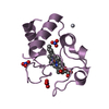
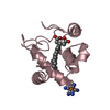
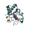
| Title | Crystal Structure of the PDZ Domain from Human RGS-3 | ||||||
 Components Components | regulator of G-protein signalling 3 isoform 1 | ||||||
 Keywords Keywords |  SIGNALING PROTEIN / PDZ Domain / RGS-3 / Human / Structural Genomics / Structural Genomics Consortium / SGC SIGNALING PROTEIN / PDZ Domain / RGS-3 / Human / Structural Genomics / Structural Genomics Consortium / SGC | ||||||
| Function / homology |  Function and homology information Function and homology informationregulation of G protein-coupled receptor signaling pathway / negative regulation of signal transduction / GTPase activator activity / G alpha (i) signalling events / G alpha (q) signalling events / G protein-coupled receptor signaling pathway / GTPase activity / nucleus / plasma membrane / cytosolSimilarity search - Function | ||||||
| Biological species |  Homo sapiens (human) Homo sapiens (human) | ||||||
| Method | X-RAY DIFFRACTION / SYNCHROTRON / MOLECULAR REPLACEMENT / Resolution: 2.39 Å | ||||||
 Authors Authors | Ugochukwu, E. / Berridge, G. / Johansson, C. / Smee, C. / Savitsky, P. / Burgess, N. / Colebrook, S. / Yang, X. / Elkins, J. / Doyle, D. ...Ugochukwu, E. / Berridge, G. / Johansson, C. / Smee, C. / Savitsky, P. / Burgess, N. / Colebrook, S. / Yang, X. / Elkins, J. / Doyle, D. / Turnbull, A. / Papagrigoriou, E. / Debreczeni, J. / Bunkoczi, G. / Gorrec, F. / von Delft, F. / Arrowsmith, C. / Sundstrom, M. / Weigelt, J. / Edwards, A. / Structural Genomics Consortium (SGC) | ||||||
 Citation Citation | Journal: To be Published Title: Crystal Structure of the PDZ Domain from Human RGS-3 Authors: Ugochukwu, E. / Berridge, G. / Johansson, C. / Smee, C. / Savitsky, P. / Burgess, N. / Colebrook, S. / Yang, X. / Elkins, J. / Doyle, D. / Turnbull, A. / Papagrigoriou, E. / Debreczeni, J. / ...Authors: Ugochukwu, E. / Berridge, G. / Johansson, C. / Smee, C. / Savitsky, P. / Burgess, N. / Colebrook, S. / Yang, X. / Elkins, J. / Doyle, D. / Turnbull, A. / Papagrigoriou, E. / Debreczeni, J. / Bunkoczi, G. / Gorrec, F. / von Delft, F. / Arrowsmith, C. / Sundstrom, M. / Weigelt, J. / Edwards, A. | ||||||
| History |
|
- Structure visualization
| Structure viewer | Molecule: MolmilJmol/JSmol |
|---|
- Downloads & links
-Download
| PDBx/mmCIF format | 2f5y.cif.gz | 48 KB | Display | PDBx/mmCIF format |
|---|---|---|---|---|
| PDB format | pdb2f5y.ent.gz | 33.3 KB | Display | PDB format |
| PDBx/mmJSON format | 2f5y.json.gz | Tree view | PDBx/mmJSON format | |
| Others |  Other downloads Other downloads |
-Validation report
| Arichive directory | https://data.pdbj.org/pub/pdb/validation_reports/f5/2f5yftp://data.pdbj.org/pub/pdb/validation_reports/f5/2f5y | HTTPS FTP |
|---|
-Related structure data


-Links
 PDBj
PDBj


- Assembly
| Deposited unit | 
| ||||||||||||||||||
|---|---|---|---|---|---|---|---|---|---|---|---|---|---|---|---|---|---|---|---|
| 1 | 
| ||||||||||||||||||
| 2 | 
| ||||||||||||||||||
| Unit cell |
| ||||||||||||||||||
| Noncrystallographic symmetry (NCS) | NCS domain:
NCS domain segments: Component-ID: 1 / Ens-ID: 1 / Beg auth comp-ID: MET / Beg label comp-ID: MET / End auth comp-ID: VAL / End label comp-ID: VAL / Refine code: 4 / Auth seq-ID: 14 - 95 / Label seq-ID: 2 - 83
|
-Components
| #1: Protein | Mass: 10239.818 Da / Num. of mol.: 2 / Fragment: PDZ Domain (residues 15-103) Source method: isolated from a genetically manipulated source Source: (gene. exp.) Homo sapiens (human) / Strain: BL21 (DE3) / Gene: RGS3 / Plasmid: pNIC28-Bsa4 / Production host:  Escherichia coli (E. coli) / References: UniProt: P49796 Escherichia coli (E. coli) / References: UniProt: P49796#2: Chemical | Sulfate  Mass: 96.063 Da / Num. of mol.: 2 / Source method: obtained synthetically / Formula: SO4 Mass: 96.063 Da / Num. of mol.: 2 / Source method: obtained synthetically / Formula: SO4#3: Water | ChemComp-HOH / | Water Mass: 18.015 Da / Num. of mol.: 42 / Source method: isolated from a natural source / Formula: H2O Mass: 18.015 Da / Num. of mol.: 42 / Source method: isolated from a natural source / Formula: H2O |
|---|
-Experimental details
-Experiment
| Experiment | Method: X-RAY DIFFRACTION / Number of used crystals: 1 |
|---|
- Sample preparation
| Crystal | Density Matthews: 2.45 Å3/Da / Density % sol: 49.71 % |
|---|---|
| Crystal grow | Temperature: 293 K / Method: vapor diffusion, sitting drop / pH: 6.5 Details: 1.6M ammonium sulphate, 0.1M MES, 10% dioxane, pH 6.5, VAPOR DIFFUSION, SITTING DROP, temperature 293K |
-Data collection
| Diffraction | Mean temperature: 100 K |
|---|---|
| Diffraction source | Source: SYNCHROTRON / Site: SLS  / Beamline: X10SA / Wavelength: 0.9788 Å / Beamline: X10SA / Wavelength: 0.9788 Å |
| Detector | Type: MARMOSAIC 225 mm CCD / Detector: CCD / Date: Oct 22, 2005 |
| Radiation | Protocol: SINGLE WAVELENGTH / Monochromatic (M) / Laue (L): M / Scattering type: x-ray |
| Radiation wavelength | Wavelength: 0.9788 Å / Relative weight: 1 |
| Reflection | Resolution: 2.39→49.86 Å / Num. obs: 8381 / % possible obs: 99.7 % / Observed criterion σ(F): 0 / Observed criterion σ(I): 0 |
| Reflection shell | Resolution: 2.4→2.49 Å / % possible all: 98.8 |
- Processing
| Software |
| |||||||||||||||||||||||||||||||||||||||||||||||||||||||||||||||||||||||||||||||||||||||||||||||||||||||||||||||||||||||||||||
|---|---|---|---|---|---|---|---|---|---|---|---|---|---|---|---|---|---|---|---|---|---|---|---|---|---|---|---|---|---|---|---|---|---|---|---|---|---|---|---|---|---|---|---|---|---|---|---|---|---|---|---|---|---|---|---|---|---|---|---|---|---|---|---|---|---|---|---|---|---|---|---|---|---|---|---|---|---|---|---|---|---|---|---|---|---|---|---|---|---|---|---|---|---|---|---|---|---|---|---|---|---|---|---|---|---|---|---|---|---|---|---|---|---|---|---|---|---|---|---|---|---|---|---|---|---|---|
| Refinement | Method to determine structure: MOLECULAR REPLACEMENT Starting model: 1I92.pdb, 1QAU.pdb Resolution: 2.39→49.88 Å / Cor.coef. Fo:Fc: 0.94 / Cor.coef. Fo:Fc free: 0.899 / SU B: 15.045 / SU ML: 0.177 / TLS residual ADP flag: LIKELY RESIDUAL / Cross valid method: THROUGHOUT / σ(F): 0 / ESU R: 0.369 / ESU R Free: 0.263 / Stereochemistry target values: MAXIMUM LIKELIHOOD / Details: HYDROGENS HAVE BEEN ADDED IN THE RIDING POSITIONS
| |||||||||||||||||||||||||||||||||||||||||||||||||||||||||||||||||||||||||||||||||||||||||||||||||||||||||||||||||||||||||||||
| Solvent computation | Ion probe radii: 0.8 Å / Shrinkage radii: 0.8 Å / VDW probe radii: 1.2 Å / Solvent model: BABINET MODEL WITH MASK | |||||||||||||||||||||||||||||||||||||||||||||||||||||||||||||||||||||||||||||||||||||||||||||||||||||||||||||||||||||||||||||
| Displacement parameters | Biso mean: 40.859 Å2
| |||||||||||||||||||||||||||||||||||||||||||||||||||||||||||||||||||||||||||||||||||||||||||||||||||||||||||||||||||||||||||||
| Refinement step | Cycle: LAST / Resolution: 2.39→49.88 Å
| |||||||||||||||||||||||||||||||||||||||||||||||||||||||||||||||||||||||||||||||||||||||||||||||||||||||||||||||||||||||||||||
| Refine LS restraints |
| |||||||||||||||||||||||||||||||||||||||||||||||||||||||||||||||||||||||||||||||||||||||||||||||||||||||||||||||||||||||||||||
| Refine LS restraints NCS | Dom-ID: 1 / Auth asym-ID: A / Ens-ID: 1 / Number: 1042 / Refine-ID: X-RAY DIFFRACTION
| |||||||||||||||||||||||||||||||||||||||||||||||||||||||||||||||||||||||||||||||||||||||||||||||||||||||||||||||||||||||||||||
| LS refinement shell | Resolution: 2.394→2.456 Å / Total num. of bins used: 20
| |||||||||||||||||||||||||||||||||||||||||||||||||||||||||||||||||||||||||||||||||||||||||||||||||||||||||||||||||||||||||||||
| Refinement TLS params. | Method: refined / Refine-ID: X-RAY DIFFRACTION
| |||||||||||||||||||||||||||||||||||||||||||||||||||||||||||||||||||||||||||||||||||||||||||||||||||||||||||||||||||||||||||||
| Refinement TLS group |
|
