 Movie
Movie Controller


+ Open data

- Basic information
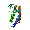
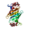
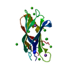
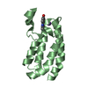
| Entry | Database: PDB / ID: 2f08 | ||||||
|---|---|---|---|---|---|---|---|
| Title | Crystal structure of a major house dust mite allergen, Derf 2 | ||||||
 Components Components | mite allergen Der f II | ||||||
 Keywords Keywords |  IMMUNE SYSTEM / immunoglobulin fold IMMUNE SYSTEM / immunoglobulin fold | ||||||
| Function / homology |  Function and homology information Function and homology information | ||||||
| Biological species |  Dermatophagoides farinae (American house dust mite) Dermatophagoides farinae (American house dust mite) | ||||||
| Method | X-RAY DIFFRACTION / MIR / Resolution: 2.2 Å | ||||||
 Authors Authors | Mikami, B. / Tanaka, Y. / Minato, N. / Suzuki, M. / Korematsu, S. | ||||||
 Citation Citation | Journal: Biochem.Biophys.Res.Commun. / Year: 2006 Title: Crystal structure and some properties of a major house dust mite allergen, Derf 2 Authors: Suzuki, M. / Tanaka, Y. / Korematsu, S. / Mikami, B. / Minato, N. | ||||||
| History |
|
- Structure visualization
| Structure viewer | Molecule: MolmilJmol/JSmol |
|---|
- Downloads & links
-Download
| PDBx/mmCIF format | 2f08.cif.gz | 106.3 KB | Display | PDBx/mmCIF format |
|---|---|---|---|---|
| PDB format | pdb2f08.ent.gz | 88.8 KB | Display | PDB format |
| PDBx/mmJSON format | 2f08.json.gz | Tree view | PDBx/mmJSON format | |
| Others |  Other downloads Other downloads |
-Validation report
| Arichive directory | https://data.pdbj.org/pub/pdb/validation_reports/f0/2f08ftp://data.pdbj.org/pub/pdb/validation_reports/f0/2f08 | HTTPS FTP |
|---|
-Related structure data
| Similar structure data |
|---|

-Links
 PDBj
PDBj
- Assembly
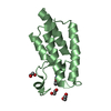
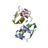
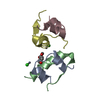
| Deposited unit | 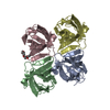
| ||||||||
|---|---|---|---|---|---|---|---|---|---|
| 1 | 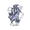
| ||||||||
| 2 | 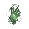
| ||||||||
| 3 | 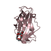
| ||||||||
| 4 | 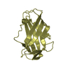
| ||||||||
| Unit cell |
| ||||||||
| Details | The biological assembly is a monomer. |
-Components
| #1: Protein | Mass: 14064.193 Da / Num. of mol.: 4 / Fragment: residues 1-129 Source method: isolated from a genetically manipulated source Source: (gene. exp.) Dermatophagoides farinae (American house dust mite)Production host:  Escherichia coli (E. coli) / Strain (production host): Rosseta(DE3)pLys / References: UniProt: Q00855 Escherichia coli (E. coli) / Strain (production host): Rosseta(DE3)pLys / References: UniProt: Q00855#2: Chemical | ChemComp-P4C / | 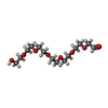  Mass: 324.367 Da / Num. of mol.: 1 / Source method: obtained synthetically / Formula: C14H28O8 Mass: 324.367 Da / Num. of mol.: 1 / Source method: obtained synthetically / Formula: C14H28O8#3: Water | ChemComp-HOH / | Water Mass: 18.015 Da / Num. of mol.: 156 / Source method: isolated from a natural source / Formula: H2O Mass: 18.015 Da / Num. of mol.: 156 / Source method: isolated from a natural source / Formula: H2O |
|---|
-Experimental details
-Experiment
| Experiment | Method: X-RAY DIFFRACTION / Number of used crystals: 1 |
|---|
- Sample preparation
| Crystal | Density Matthews: 2.62 Å3/Da / Density % sol: 53.06 % |
|---|---|
| Crystal grow | Temperature: 293 K / Method: vapor diffusion, hanging drop / pH: 7 Details: 23% PEG 4000, 0.06M ammonium sulfate, 0.1M sodium acetate, 10mM HEPES , pH 7.0, VAPOR DIFFUSION, HANGING DROP, temperature 293K |
-Data collection
| Diffraction | Mean temperature: 298 K |
|---|---|
| Diffraction source | Source: ROTATING ANODE / Type: MACSCIENCE / Wavelength: 1.54 Å |
| Detector | Type: SIEMENS HI-STAR / Detector: AREA DETECTOR / Date: Dec 21, 1999 / Details: carbon monochrometer |
| Radiation | Monochromator: carbon monochrometer / Protocol: SINGLE WAVELENGTH / Monochromatic (M) / Laue (L): M / Scattering type: x-ray |
| Radiation wavelength | Wavelength: 1.54 Å / Relative weight: 1 |
| Reflection | Resolution: 2.1→28.3 Å / Num. all: 34340 / Num. obs: 32658 / % possible obs: 95.05 % / Observed criterion σ(I): 1 / Redundancy: 3.38 % / Biso Wilson estimate: 12.1 Å2 / Rsym value: 0.058 / Net I/σ(I): 25.83 |
| Reflection shell | Resolution: 2.1→2.175 Å / Redundancy: 3.37 % / Mean I/σ(I) obs: 2.61 / Num. unique all: 3009 / Rsym value: 0.342 / % possible all: 99.1 |
- Processing
| Software |
| ||||||||||||||||||||||||||||||||||||
|---|---|---|---|---|---|---|---|---|---|---|---|---|---|---|---|---|---|---|---|---|---|---|---|---|---|---|---|---|---|---|---|---|---|---|---|---|---|
| Refinement | Method to determine structure: MIR / Resolution: 2.2→14.91 Å / Rfactor Rfree error: 0.005 / Data cutoff high absF: 942193.23 / Data cutoff low absF: 0 / Isotropic thermal model: RESTRAINED / Cross valid method: THROUGHOUT / σ(F): 0 / Stereochemistry target values: Engh & Huber
| ||||||||||||||||||||||||||||||||||||
| Solvent computation | Solvent model: FLAT MODEL / Bsol: 39.6915 Å2 / ksol: 0.259142 e/Å3 | ||||||||||||||||||||||||||||||||||||
| Displacement parameters | Biso mean: 35.5 Å2
| ||||||||||||||||||||||||||||||||||||
| Refine analyze |
| ||||||||||||||||||||||||||||||||||||
| Refinement step | Cycle: LAST / Resolution: 2.2→14.91 Å
| ||||||||||||||||||||||||||||||||||||
| Refine LS restraints |
| ||||||||||||||||||||||||||||||||||||
| LS refinement shell | Resolution: 2.2→2.28 Å / Rfactor Rfree error: 0.017 / Total num. of bins used: 6
| ||||||||||||||||||||||||||||||||||||
| Xplor file |
|
