 Movie
Movie Controller

[English] 日本語
 Yorodumi
- PDB-2exx: Crystal structure of HSCARG from Homo sapiens in complex with NADP -
+ Open data

- Basic information
| Entry | Database: PDB / ID: 2exx | ||||||
|---|---|---|---|---|---|---|---|
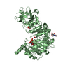
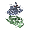
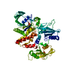
| Title | Crystal structure of HSCARG from Homo sapiens in complex with NADP | ||||||
 Components Components | HSCARG protein | ||||||
 Keywords Keywords | UNKNOWN FUNCTION / Protein-NADP complex | ||||||
| Function / homology |  Function and homology information Function and homology information Urea cycle / perinuclear region of cytoplasm / nucleoplasm / identical protein binding / nucleus / cytosol Urea cycle / perinuclear region of cytoplasm / nucleoplasm / identical protein binding / nucleus / cytosolSimilarity search - Function | ||||||
| Biological species |  Homo sapiens (human) Homo sapiens (human) | ||||||
| Method | X-RAY DIFFRACTION / SYNCHROTRON / SAD / Resolution: 2.4 Å | ||||||
 Authors Authors | Dai, X. / Chen, Q. / Yao, D. / Liang, Y. / Dong, Y. / Gu, X. / Zheng, X. / Luo, M. | ||||||
 Citation Citation | Journal: Proc.Natl.Acad.Sci.USA / Year: 2007 Title: Restructuring of the dinucleotide-binding fold in an NADP(H) sensor protein. Authors: Zheng, X. / Dai, X. / Zhao, Y. / Chen, Q. / Lu, F. / Yao, D. / Yu, Q. / Liu, X. / Zhang, C. / Gu, X. / Luo, M. | ||||||
| History |
|
- Structure visualization
| Structure viewer | Molecule: MolmilJmol/JSmol |
|---|
- Downloads & links
-Download
| PDBx/mmCIF format | 2exx.cif.gz | 122.9 KB | Display | PDBx/mmCIF format |
|---|---|---|---|---|
| PDB format | pdb2exx.ent.gz | 102.5 KB | Display | PDB format |
| PDBx/mmJSON format | 2exx.json.gz | Tree view | PDBx/mmJSON format | |
| Others |  Other downloads Other downloads |
-Validation report
| Arichive directory | https://data.pdbj.org/pub/pdb/validation_reports/ex/2exxftp://data.pdbj.org/pub/pdb/validation_reports/ex/2exx | HTTPS FTP |
|---|
-Related structure data
| Similar structure data |
|---|


-Links
 PDBj
PDBj
- Assembly
| Deposited unit | 
| ||||||||
|---|---|---|---|---|---|---|---|---|---|
| 1 |
| ||||||||
| Unit cell |
| ||||||||
| Details | The biological assembly is a dimer generated from the two molecules in the asymmetric unit |
-Components
| #1: Protein | Mass: 34165.348 Da / Num. of mol.: 2 Source method: isolated from a genetically manipulated source Source: (gene. exp.) Homo sapiens (human) / Production host:  Escherichia coli (E. coli) / References: GenBank: 13938446, UniProt: Q9HBL8*PLUS Escherichia coli (E. coli) / References: GenBank: 13938446, UniProt: Q9HBL8*PLUS#2: Chemical | ChemComp-NAP / | Nicotinamide adenine dinucleotide phosphate  Mass: 743.405 Da / Num. of mol.: 1 / Source method: obtained synthetically / Formula: C21H28N7O17P3 Mass: 743.405 Da / Num. of mol.: 1 / Source method: obtained synthetically / Formula: C21H28N7O17P3#3: Chemical | ChemComp-GOL / | Glycerol  Mass: 92.094 Da / Num. of mol.: 1 / Source method: obtained synthetically / Formula: C3H8O3 Mass: 92.094 Da / Num. of mol.: 1 / Source method: obtained synthetically / Formula: C3H8O3#4: Water | ChemComp-HOH / | Water Mass: 18.015 Da / Num. of mol.: 90 / Source method: isolated from a natural source / Formula: H2O Mass: 18.015 Da / Num. of mol.: 90 / Source method: isolated from a natural source / Formula: H2O |
|---|
-Experimental details
-Experiment
| Experiment | Method: X-RAY DIFFRACTION / Number of used crystals: 1 |
|---|
- Sample preparation
| Crystal | Density Matthews: 3.39 Å3/Da / Density % sol: 63.75 % |
|---|---|
| Crystal grow | Temperature: 277 K / Method: vapor diffusion, hanging drop / pH: 7 Details: 1.2M sodium potassium phosphate, pH 7.0, VAPOR DIFFUSION, HANGING DROP, temperature 277K |
-Data collection
| Diffraction | Mean temperature: 100 K |
|---|---|
| Diffraction source | Source: SYNCHROTRON / Site: APS  / Beamline: 22-BM / Wavelength: 0.97928 Å / Beamline: 22-BM / Wavelength: 0.97928 Å |
| Detector | Type: MARMOSAIC 225 mm CCD / Detector: CCD / Date: Aug 6, 2005 |
| Radiation | Monochromator: Si 111 cut crystal / Protocol: SINGLE WAVELENGTH / Monochromatic (M) / Laue (L): M / Scattering type: x-ray |
| Radiation wavelength | Wavelength: 0.97928 Å / Relative weight: 1 |
| Reflection | Resolution: 2.4→50 Å / Num. all: 35699 / Num. obs: 34021 / % possible obs: 95.3 % / Observed criterion σ(I): 3 |
| Reflection shell | Resolution: 2.4→2.49 Å / % possible all: 100 |
- Processing
| Software |
| ||||||||||||||||||||
|---|---|---|---|---|---|---|---|---|---|---|---|---|---|---|---|---|---|---|---|---|---|
| Refinement | Method to determine structure: SAD / Resolution: 2.4→30 Å / σ(F): 0 / Stereochemistry target values: Engh & Huber
| ||||||||||||||||||||
| Refinement step | Cycle: LAST / Resolution: 2.4→30 Å
|
