 Movie
Movie Controller


+ Open data

- Basic information
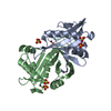
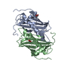
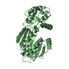
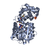
| Entry | Database: PDB / ID: 2eui | ||||||
|---|---|---|---|---|---|---|---|
| Title | Crystal structure of a probable acetyltransferase | ||||||
 Components Components | Probable acetyltransferase | ||||||
 Keywords Keywords |  STRUCTURAL GENOMICS / UNKNOWN FUNCTION / transferase / dimer / T1065 / PSI / Protein Structure Initiative / New York SGX Research Center for Structural Genomics / NYSGXRC STRUCTURAL GENOMICS / UNKNOWN FUNCTION / transferase / dimer / T1065 / PSI / Protein Structure Initiative / New York SGX Research Center for Structural Genomics / NYSGXRC | ||||||
| Function / homology |  Function and homology informationN-acetyltransferase activity / acyltransferase activity, transferring groups other than amino-acyl groups Function and homology informationN-acetyltransferase activity / acyltransferase activity, transferring groups other than amino-acyl groupsSimilarity search - Function | ||||||
| Biological species |  Pseudomonas aeruginosa PAO1 (bacteria) Pseudomonas aeruginosa PAO1 (bacteria) | ||||||
| Method | X-RAY DIFFRACTION / SYNCHROTRON / MAD / Resolution: 2.8 Å | ||||||
 Authors Authors | Swaminathan, S. / Eswaramoorthy, S. / Burley, S.K. / New York SGX Research Center for Structural Genomics (NYSGXRC) | ||||||
 Citation Citation | Journal: To be Published Title: Crystal structure of a probable acetyltransferase Authors: Eswaramoorthy, S. / Swaminathan, S. | ||||||
| History |
|
- Structure visualization
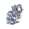
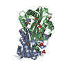
| Structure viewer | Molecule: MolmilJmol/JSmol |
|---|
- Downloads & links
-Download
| PDBx/mmCIF format | 2eui.cif.gz | 128.7 KB | Display | PDBx/mmCIF format |
|---|---|---|---|---|
| PDB format | pdb2eui.ent.gz | 109.1 KB | Display | PDB format |
| PDBx/mmJSON format | 2eui.json.gz | Tree view | PDBx/mmJSON format | |
| Others |  Other downloads Other downloads |
-Validation report
| Arichive directory | https://data.pdbj.org/pub/pdb/validation_reports/eu/2euiftp://data.pdbj.org/pub/pdb/validation_reports/eu/2eui | HTTPS FTP |
|---|
-Related structure data
| Similar structure data | |
|---|---|
| Other databases |


-Links
 PDBj
PDBj

- Assembly
| Deposited unit | 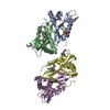
| ||||||||
|---|---|---|---|---|---|---|---|---|---|
| 1 | 
| ||||||||
| 2 | 
| ||||||||
| Unit cell |
|
-Components
| #1: Protein | Mass: 18262.301 Da / Num. of mol.: 4 Source method: isolated from a genetically manipulated source Source: (gene. exp.) Pseudomonas aeruginosa PAO1 (bacteria) / Species: Pseudomonas aeruginosa / Strain: PA01 / Production host: Escherichia coli (E. coli) / References: UniProt: Q9HX01#2: Water | ChemComp-HOH / | Water Mass: 18.015 Da / Num. of mol.: 137 / Source method: isolated from a natural source / Formula: H2O Mass: 18.015 Da / Num. of mol.: 137 / Source method: isolated from a natural source / Formula: H2O |
|---|
-Experimental details
-Experiment
| Experiment | Method: X-RAY DIFFRACTION / Number of used crystals: 1 |
|---|
- Sample preparation
| Crystal | Density Matthews: 3.03 Å3/Da / Density % sol: 59.41 % |
|---|---|
| Crystal grow | Temperature: 291 K / Method: vapor diffusion, sitting drop / pH: 5.5 Details: 25% PEG 3350, 0.2M Magnesium Chloride, pH 5.5, VAPOR DIFFUSION, SITTING DROP, temperature 291K |
-Data collection
| Diffraction | Mean temperature: 100 K | ||||||||||||
|---|---|---|---|---|---|---|---|---|---|---|---|---|---|
| Diffraction source | Source: SYNCHROTRON / Site: NSLS  / Beamline: X25 / Wavelength: 0.9805, 0.9802, 0.9400 / Beamline: X25 / Wavelength: 0.9805, 0.9802, 0.9400 | ||||||||||||
| Detector | Type: ADSC QUANTUM 315 / Detector: CCD / Date: Sep 28, 2005 | ||||||||||||
| Radiation | Monochromator: GRAPHITE / Protocol: MAD / Monochromatic (M) / Laue (L): M / Scattering type: x-ray | ||||||||||||
| Radiation wavelength |
| ||||||||||||
| Reflection | Resolution: 2.8→50 Å / Num. all: 21842 / Num. obs: 21842 / % possible obs: 100 % / Observed criterion σ(F): 0 / Observed criterion σ(I): 0 / Redundancy: 13.6 % / Rmerge(I) obs: 0.139 / Net I/σ(I): 6.6 | ||||||||||||
| Reflection shell | Resolution: 2.8→2.9 Å / Redundancy: 9.7 % / Rmerge(I) obs: 0.473 / Num. unique all: 2173 / % possible all: 99.7 |
- Processing
| Software |
| |||||||||||||||||||||||||
|---|---|---|---|---|---|---|---|---|---|---|---|---|---|---|---|---|---|---|---|---|---|---|---|---|---|---|
| Refinement | Method to determine structure: MAD / Resolution: 2.8→50 Å / Cross valid method: THROUGHOUT / σ(F): 0 / Stereochemistry target values: Engh & Huber
| |||||||||||||||||||||||||
| Refinement step | Cycle: LAST / Resolution: 2.8→50 Å
| |||||||||||||||||||||||||
| Refine LS restraints |
|
