 Movie
Movie Controller

[English] 日本語
 Yorodumi
- PDB-2es7: Crystal structure of Q8ZP25 from Salmonella typhimurium LT2. NESG... -
+ Open data

- Basic information
| Entry | Database: PDB / ID: 2es7 | ||||||
|---|---|---|---|---|---|---|---|
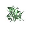
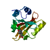
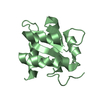
| Title | Crystal structure of Q8ZP25 from Salmonella typhimurium LT2. NESG TARGET STR70 | ||||||
 Components Components | putative thiol-disulfide isomerase and thioredoxin | ||||||
 Keywords Keywords |  ISOMERASE / STRUCTURAL GENOMICS / PSI / PROTEIN STRUCTURE INITIATIVE / NORTHEAST STRUCTURAL GENOMICS CONSORTIUM / NESG ISOMERASE / STRUCTURAL GENOMICS / PSI / PROTEIN STRUCTURE INITIATIVE / NORTHEAST STRUCTURAL GENOMICS CONSORTIUM / NESG | ||||||
| Function / homology |  Function and homology information Function and homology information | ||||||
| Biological species |  Salmonella typhimurium (bacteria) Salmonella typhimurium (bacteria) | ||||||
| Method | X-RAY DIFFRACTION / SYNCHROTRON / MAD / Resolution: 2.8 Å | ||||||
 Authors Authors | Benach, J. / Su, M. / Forouhar, F. / Chen, Y. / Ho, C.K. / Janjua, H. / Cunningham, K. / Ma, L.-C. / Rong, X. / Liu, J. ...Benach, J. / Su, M. / Forouhar, F. / Chen, Y. / Ho, C.K. / Janjua, H. / Cunningham, K. / Ma, L.-C. / Rong, X. / Liu, J. / Baran, M. / Acton, T.B. / Rost, B. / Montelione, G.T. / Tong, L. / Hunt, J.F. / Northeast Structural Genomics Consortium (NESG) | ||||||
 Citation Citation | Journal: To be Published Title: Crystal structure of Q8ZP25 from Salmonella typhimurium LT2 NESG target STR70. Authors: Benach, J. / Su, M. / Forouhar, F. / Chen, Y. / Janjua, H. / Rong, B.X. / Acton, T.B. / Montelione, G.T. / Hunt, J.F. / Tong, L. | ||||||
| History |
|
- Structure visualization
| Structure viewer | Molecule: MolmilJmol/JSmol |
|---|
- Downloads & links
-Download
| PDBx/mmCIF format | 2es7.cif.gz | 105.1 KB | Display | PDBx/mmCIF format |
|---|---|---|---|---|
| PDB format | pdb2es7.ent.gz | 85.4 KB | Display | PDB format |
| PDBx/mmJSON format | 2es7.json.gz | Tree view | PDBx/mmJSON format | |
| Others |  Other downloads Other downloads |
-Validation report
| Arichive directory | https://data.pdbj.org/pub/pdb/validation_reports/es/2es7ftp://data.pdbj.org/pub/pdb/validation_reports/es/2es7 | HTTPS FTP |
|---|
-Related structure data
| Similar structure data | |
|---|---|
| Other databases |


-Links
 PDBj
PDBj- Assembly
| Deposited unit | 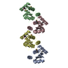
| ||||||||
|---|---|---|---|---|---|---|---|---|---|
| 1 | 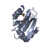
| ||||||||
| 2 | 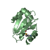
| ||||||||
| 3 | 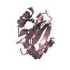
| ||||||||
| 4 | 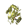
| ||||||||
| Unit cell |
|
-Components
| #1: Protein | Mass: 16286.870 Da / Num. of mol.: 4 Source method: isolated from a genetically manipulated source Source: (gene. exp.) Salmonella typhimurium (bacteria) / Strain: LT2 / Gene: STM1790 / Plasmid: pET21 / Production host: Escherichia coli (E. coli) / References: GenBank: 16420320, UniProt: Q8ZP25*PLUS#2: Water | ChemComp-HOH / | Water Mass: 18.015 Da / Num. of mol.: 560 / Source method: isolated from a natural source / Formula: H2O Mass: 18.015 Da / Num. of mol.: 560 / Source method: isolated from a natural source / Formula: H2O |
|---|
-Experimental details
-Experiment
| Experiment | Method: X-RAY DIFFRACTION / Number of used crystals: 1 |
|---|
- Sample preparation
| Crystal | Density Matthews: 2.07 Å3/Da / Density % sol: 40.6 % |
|---|---|
| Crystal grow | Method: vapor diffusion, hanging drop / Details: VAPOR DIFFUSION, HANGING DROP |
-Data collection
| Diffraction | Mean temperature: 100 K | ||||||||||||
|---|---|---|---|---|---|---|---|---|---|---|---|---|---|
| Diffraction source | Source: SYNCHROTRON / Site: NSLS  / Beamline: X4A / Wavelength: 0.97925, 0.97955, 0.96782 / Beamline: X4A / Wavelength: 0.97925, 0.97955, 0.96782 | ||||||||||||
| Detector | Type: ADSC QUANTUM 4 / Detector: CCD / Date: Aug 1, 2005 | ||||||||||||
| Radiation | Monochromator: SI 111 CHANNEL / Protocol: MAD / Monochromatic (M) / Laue (L): M / Scattering type: x-ray | ||||||||||||
| Radiation wavelength |
| ||||||||||||
| Reflection | Resolution: 2.8→20 Å / Num. obs: 25130 / % possible obs: 98.9 % / Observed criterion σ(I): -3 / Redundancy: 4 % / Biso Wilson estimate: 85.1 Å2 / Rmerge(I) obs: 0.049 / Net I/σ(I): 35.37 | ||||||||||||
| Reflection shell | Resolution: 2.8→2.85 Å / Redundancy: 3.4 % / Rmerge(I) obs: 0.122 / Mean I/σ(I) obs: 9.5 / % possible all: 97.4 |
- Processing
| Software |
| ||||||||||||||||||||||||||||||||||||||||||||||||||||||||||||||||||||||||||||||||
|---|---|---|---|---|---|---|---|---|---|---|---|---|---|---|---|---|---|---|---|---|---|---|---|---|---|---|---|---|---|---|---|---|---|---|---|---|---|---|---|---|---|---|---|---|---|---|---|---|---|---|---|---|---|---|---|---|---|---|---|---|---|---|---|---|---|---|---|---|---|---|---|---|---|---|---|---|---|---|---|---|---|
| Refinement | Method to determine structure: MAD / Resolution: 2.8→19.9 Å / Rfactor Rfree error: 0.007 / Data cutoff high absF: 364563.16 / Data cutoff low absF: 0 / Isotropic thermal model: RESTRAINED / Cross valid method: THROUGHOUT / σ(F): 2
| ||||||||||||||||||||||||||||||||||||||||||||||||||||||||||||||||||||||||||||||||
| Solvent computation | Solvent model: FLAT MODEL / Bsol: 98.4149 Å2 / ksol: 0.159752 e/Å3 | ||||||||||||||||||||||||||||||||||||||||||||||||||||||||||||||||||||||||||||||||
| Displacement parameters | Biso mean: 36.2 Å2
| ||||||||||||||||||||||||||||||||||||||||||||||||||||||||||||||||||||||||||||||||
| Refine analyze |
| ||||||||||||||||||||||||||||||||||||||||||||||||||||||||||||||||||||||||||||||||
| Refinement step | Cycle: LAST / Resolution: 2.8→19.9 Å
| ||||||||||||||||||||||||||||||||||||||||||||||||||||||||||||||||||||||||||||||||
| Refine LS restraints |
| ||||||||||||||||||||||||||||||||||||||||||||||||||||||||||||||||||||||||||||||||
| LS refinement shell | Resolution: 2.8→2.97 Å / Rfactor Rfree error: 0.02 / Total num. of bins used: 6
| ||||||||||||||||||||||||||||||||||||||||||||||||||||||||||||||||||||||||||||||||
| Xplor file |
|
