 Movie
Movie Controller


+ Open data

- Basic information
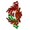
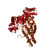
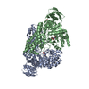
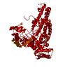
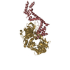
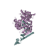
| Entry | Database: PDB / ID: 2epo | ||||||
|---|---|---|---|---|---|---|---|
| Title | N-acetyl-B-D-glucosaminidase (GCNA) from Streptococcus gordonii | ||||||
 Components Components | N-acetyl-beta-D-glucosaminidase | ||||||
 Keywords Keywords |  HYDROLASE / glycoside hydrolase / family 20 / gcna / glucosaminidase HYDROLASE / glycoside hydrolase / family 20 / gcna / glucosaminidase | ||||||
| Function / homology |  Function and homology informationbeta-N-acetylhexosaminidase activity / beta-N-acetylhexosaminidase / N-acetyl-beta-D-galactosaminidase activity / metabolic process Function and homology informationbeta-N-acetylhexosaminidase activity / beta-N-acetylhexosaminidase / N-acetyl-beta-D-galactosaminidase activity / metabolic processSimilarity search - Function | ||||||
| Biological species |  Streptococcus gordonii (bacteria) Streptococcus gordonii (bacteria) | ||||||
| Method | X-RAY DIFFRACTION / SYNCHROTRON / MOLECULAR REPLACEMENT / Resolution: 1.56 Å | ||||||
 Authors Authors | Langley, D.B. | ||||||
 Citation Citation | Journal: J.Mol.Biol. / Year: 2008 Title: Structure of N-acetyl-beta-D-glucosaminidase (GcnA) from the Endocarditis Pathogen Streptococcus gordonii and its Complex with the Mechanism-based Inhibitor NAG-thiazoline Authors: Langley, D.B. / Harty, D.W.S. / Jacques, N.A. / Hunter, N. / Guss, J.M. / Collyer, C.A. | ||||||
| History |
|
- Structure visualization
| Structure viewer | Molecule: MolmilJmol/JSmol |
|---|
- Downloads & links
-Download
| PDBx/mmCIF format | 2epo.cif.gz | 263.5 KB | Display | PDBx/mmCIF format |
|---|---|---|---|---|
| PDB format | pdb2epo.ent.gz | 211.5 KB | Display | PDB format |
| PDBx/mmJSON format | 2epo.json.gz | Tree view | PDBx/mmJSON format | |
| Others |  Other downloads Other downloads |
-Validation report
| Arichive directory | https://data.pdbj.org/pub/pdb/validation_reports/ep/2epoftp://data.pdbj.org/pub/pdb/validation_reports/ep/2epo | HTTPS FTP |
|---|
-Related structure data


-Links
 PDBj
PDBj

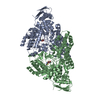
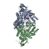
- Assembly
| Deposited unit | 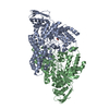
| ||||||||
|---|---|---|---|---|---|---|---|---|---|
| 1 |
| ||||||||
| Unit cell |
| ||||||||
| Components on special symmetry positions |
|
-Components
| #1: Protein | Mass: 72331.547 Da / Num. of mol.: 2 Source method: isolated from a genetically manipulated source Source: (gene. exp.) Streptococcus gordonii (bacteria) / Strain: FSS2 / Gene: gcna / Plasmid: pHAR101 / Production host: Escherichia coli (E. coli) / References: UniProt: Q6ST21, beta-N-acetylhexosaminidase#2: Chemical | Acetic acid  Mass: 60.052 Da / Num. of mol.: 2 / Source method: obtained synthetically / Formula: C2H4O2 Mass: 60.052 Da / Num. of mol.: 2 / Source method: obtained synthetically / Formula: C2H4O2#3: Water | ChemComp-HOH / | Water Mass: 18.015 Da / Num. of mol.: 605 / Source method: isolated from a natural source / Formula: H2O Mass: 18.015 Da / Num. of mol.: 605 / Source method: isolated from a natural source / Formula: H2O |
|---|
-Experimental details
-Experiment
| Experiment | Method: X-RAY DIFFRACTION / Number of used crystals: 1 |
|---|
- Sample preparation
| Crystal | Density Matthews: 2.22 Å3/Da / Density % sol: 44.7 % |
|---|---|
| Crystal grow | Temperature: 293 K / Method: vapor diffusion, hanging drop / pH: 6 Details: 100mM sodium citrate, 200mM ammonium acetate, 24%(v/v) PEG4000, pH 6.0, VAPOR DIFFUSION, HANGING DROP, temperature 293K |
-Data collection
| Diffraction | Mean temperature: 100 K |
|---|---|
| Diffraction source | Source: SYNCHROTRON / Site: MAX II  / Beamline: I711 / Wavelength: 1.008 Å / Beamline: I711 / Wavelength: 1.008 Å |
| Detector | Type: MAR CCD 165 mm / Detector: CCD / Date: Feb 1, 2004 |
| Radiation | Monochromator: single crystal of Si(111) / Protocol: SINGLE WAVELENGTH / Monochromatic (M) / Laue (L): M / Scattering type: x-ray |
| Radiation wavelength | Wavelength: 1.008 Å / Relative weight: 1 |
| Reflection | Resolution: 1.55→34.88 Å / Num. all: 184801 / Num. obs: 170106 / % possible obs: 96.85 % / Observed criterion σ(F): 3 / Observed criterion σ(I): 3 / Redundancy: 3.6 % / Biso Wilson estimate: 19.9 Å2 / Rmerge(I) obs: 0.093 / Net I/σ(I): 11.7 |
| Reflection shell | Resolution: 1.55→1.6 Å / Redundancy: 4.3 % / Rmerge(I) obs: 0.43 / Mean I/σ(I) obs: 4.36 / % possible all: 77.6 |
- Processing
| Software |
| |||||||||||||||||||||||||||||||||||||||||||||||||||||||||||||||||||||||||||||||||||||||||||||||||||||||||||||||||||||||||||||
|---|---|---|---|---|---|---|---|---|---|---|---|---|---|---|---|---|---|---|---|---|---|---|---|---|---|---|---|---|---|---|---|---|---|---|---|---|---|---|---|---|---|---|---|---|---|---|---|---|---|---|---|---|---|---|---|---|---|---|---|---|---|---|---|---|---|---|---|---|---|---|---|---|---|---|---|---|---|---|---|---|---|---|---|---|---|---|---|---|---|---|---|---|---|---|---|---|---|---|---|---|---|---|---|---|---|---|---|---|---|---|---|---|---|---|---|---|---|---|---|---|---|---|---|---|---|---|
| Refinement | Method to determine structure: MOLECULAR REPLACEMENT Starting model: Selenium methionine structure Resolution: 1.56→34.88 Å / Cor.coef. Fo:Fc: 0.948 / Cor.coef. Fo:Fc free: 0.922 / SU B: 2.073 / SU ML: 0.074 / Cross valid method: THROUGHOUT / σ(F): 0 / σ(I): 0 / ESU R: 0.097 / ESU R Free: 0.1 / Stereochemistry target values: MAXIMUM LIKELIHOOD / Details: HYDROGENS HAVE BEEN ADDED IN THE RIDING POSITIONS
| |||||||||||||||||||||||||||||||||||||||||||||||||||||||||||||||||||||||||||||||||||||||||||||||||||||||||||||||||||||||||||||
| Solvent computation | Solvent model: BABINET MODEL PARAMETERS FOR MASK CACLULATION | |||||||||||||||||||||||||||||||||||||||||||||||||||||||||||||||||||||||||||||||||||||||||||||||||||||||||||||||||||||||||||||
| Displacement parameters | Biso mean: 19.128 Å2
| |||||||||||||||||||||||||||||||||||||||||||||||||||||||||||||||||||||||||||||||||||||||||||||||||||||||||||||||||||||||||||||
| Refine analyze | Luzzati coordinate error obs: 0.074 Å | |||||||||||||||||||||||||||||||||||||||||||||||||||||||||||||||||||||||||||||||||||||||||||||||||||||||||||||||||||||||||||||
| Refinement step | Cycle: LAST / Resolution: 1.56→34.88 Å
| |||||||||||||||||||||||||||||||||||||||||||||||||||||||||||||||||||||||||||||||||||||||||||||||||||||||||||||||||||||||||||||
| Refine LS restraints |
| |||||||||||||||||||||||||||||||||||||||||||||||||||||||||||||||||||||||||||||||||||||||||||||||||||||||||||||||||||||||||||||
| LS refinement shell | Resolution: 1.555→1.596 Å / Total num. of bins used: 20
|
