 Movie
Movie Controller

[English] 日本語
 Yorodumi
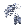
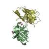
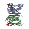
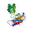
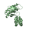
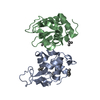
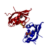
- PDB-2eg9: Crystal structure of the truncated extracellular domain of mouse CD38 -
+ Open data

- Basic information
| Entry | Database: PDB / ID: 2eg9 | ||||||
|---|---|---|---|---|---|---|---|
| Title | Crystal structure of the truncated extracellular domain of mouse CD38 | ||||||
 Components Components | ADP-ribosyl cyclase 1 Cyclic ADP-ribose Cyclic ADP-ribose | ||||||
 Keywords Keywords | HYDROLASE / CELL SUEFACE ANTIGEN / Structural Genomics / NPPSFA / National Project on Protein Structural and Functional Analyses / RIKEN Structural Genomics/Proteomics Initiative / RSGI | ||||||
| Function / homology |  Function and homology informationNicotinate metabolism / 2'-phospho-ADP-ribosyl cyclase/2'-phospho-cyclic-ADP-ribose transferase / phosphorus-oxygen lyase activity / response to xenobiotic stimulus => GO:0009410 / artery smooth muscle contraction / NADP+ nucleosidase activity / NAD+ nucleosidase activity / ADP-ribosyl cyclase/cyclic ADP-ribose hydrolase / NAD+ nucleotidase, cyclic ADP-ribose generating / hydrolase activity, acting on glycosyl bonds ...Nicotinate metabolism / 2'-phospho-ADP-ribosyl cyclase/2'-phospho-cyclic-ADP-ribose transferase / phosphorus-oxygen lyase activity / response to xenobiotic stimulus => GO:0009410 / artery smooth muscle contraction / NADP+ nucleosidase activity / NAD+ nucleosidase activity / ADP-ribosyl cyclase/cyclic ADP-ribose hydrolase / NAD+ nucleotidase, cyclic ADP-ribose generating / hydrolase activity, acting on glycosyl bonds / negative regulation of bone resorption / response to hydroperoxide / long-term synaptic depression / response to retinoic acid / positive regulation of B cell proliferation / positive regulation of vasoconstriction / response to interleukin-1 / secretory granule membrane / response to progesterone / response to hormone / female pregnancy / B cell receptor signaling pathway / positive regulation of insulin secretion / response to estradiol / negative regulation of neuron projection development / positive regulation of cytosolic calcium ion concentration / transferase activity / positive regulation of cell growth / basolateral plasma membrane / membrane => GO:0016020 / response to hypoxia / intracellular membrane-bounded organelle / negative regulation of DNA-templated transcription / positive regulation of cell population proliferation / negative regulation of apoptotic process / positive regulation of DNA-templated transcription / cell surface / membrane / identical protein binding / nucleus / plasma membrane Function and homology informationNicotinate metabolism / 2'-phospho-ADP-ribosyl cyclase/2'-phospho-cyclic-ADP-ribose transferase / phosphorus-oxygen lyase activity / response to xenobiotic stimulus => GO:0009410 / artery smooth muscle contraction / NADP+ nucleosidase activity / NAD+ nucleosidase activity / ADP-ribosyl cyclase/cyclic ADP-ribose hydrolase / NAD+ nucleotidase, cyclic ADP-ribose generating / hydrolase activity, acting on glycosyl bonds ...Nicotinate metabolism / 2'-phospho-ADP-ribosyl cyclase/2'-phospho-cyclic-ADP-ribose transferase / phosphorus-oxygen lyase activity / response to xenobiotic stimulus => GO:0009410 / artery smooth muscle contraction / NADP+ nucleosidase activity / NAD+ nucleosidase activity / ADP-ribosyl cyclase/cyclic ADP-ribose hydrolase / NAD+ nucleotidase, cyclic ADP-ribose generating / hydrolase activity, acting on glycosyl bonds / negative regulation of bone resorption / response to hydroperoxide / long-term synaptic depression / response to retinoic acid / positive regulation of B cell proliferation / positive regulation of vasoconstriction / response to interleukin-1 / secretory granule membrane / response to progesterone / response to hormone / female pregnancy / B cell receptor signaling pathway / positive regulation of insulin secretion / response to estradiol / negative regulation of neuron projection development / positive regulation of cytosolic calcium ion concentration / transferase activity / positive regulation of cell growth / basolateral plasma membrane / membrane => GO:0016020 / response to hypoxia / intracellular membrane-bounded organelle / negative regulation of DNA-templated transcription / positive regulation of cell population proliferation / negative regulation of apoptotic process / positive regulation of DNA-templated transcription / cell surface / membrane / identical protein binding / nucleus / plasma membraneSimilarity search - Function | ||||||
| Biological species |  Mus musculus (house mouse) Mus musculus (house mouse) | ||||||
| Method | X-RAY DIFFRACTION / SYNCHROTRON / MOLECULAR REPLACEMENT / Resolution: 2.8 Å | ||||||
 Authors Authors | Kukimoto-Niino, M. / Mishima, C. / Wakiyama, M. / Terada, T. / Shirouzu, M. / Hara-Yokoyama, M. / Yokoyama, S. / RIKEN Structural Genomics/Proteomics Initiative (RSGI) | ||||||
 Citation Citation | Journal: To be Published Title: Crystal structure of the truncated extracellular domain of mouse CD38 Authors: Kukimoto-Niino, M. / Mishima, C. / Wakiyama, M. / Terada, T. / Shirouzu, M. / Hara-Yokoyama, M. / Yokoyama, S. | ||||||
| History |
|
- Structure visualization
| Structure viewer | Molecule: MolmilJmol/JSmol |
|---|
- Downloads & links
-Download
| PDBx/mmCIF format | 2eg9.cif.gz | 97 KB | Display | PDBx/mmCIF format |
|---|---|---|---|---|
| PDB format | pdb2eg9.ent.gz | 73.4 KB | Display | PDB format |
| PDBx/mmJSON format | 2eg9.json.gz | Tree view | PDBx/mmJSON format | |
| Others |  Other downloads Other downloads |
-Validation report
| Arichive directory | https://data.pdbj.org/pub/pdb/validation_reports/eg/2eg9ftp://data.pdbj.org/pub/pdb/validation_reports/eg/2eg9 | HTTPS FTP |
|---|
-Related structure data
| Related structure data |  2df1 S: Starting model for refinement |
|---|---|
| Similar structure data | |
| Other databases |


-Links
 PDBj
PDBj
- Assembly
| Deposited unit | 
| ||||||||
|---|---|---|---|---|---|---|---|---|---|
| 1 | 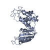
| ||||||||
| 2 | 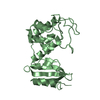
| ||||||||
| Unit cell |
|
-Components
| #1: Protein | Cyclic ADP-ribose / CD38 / Cyclic ADP-ribose hydrolase 1 / cADPr hydrolase 1 / NIM-R5 antigen / I-19 Mass: 29148.295 Da / Num. of mol.: 2 / Fragment: extracellular domain (residues 48-288) / Mutation: N104D, N124D, N213D, N223D Source method: isolated from a genetically manipulated source Source: (gene. exp.) Mus musculus (house mouse) / Gene: Cd38 / Plasmid: pAc-BiP / Cell line (production host): Schneider Line 2 (S2) / Production host:  Drosophila melanogaster (fruit fly) / References: UniProt: P56528, NAD+ glycohydrolase Drosophila melanogaster (fruit fly) / References: UniProt: P56528, NAD+ glycohydrolase#2: Water | ChemComp-HOH / | Water Mass: 18.015 Da / Num. of mol.: 23 / Source method: isolated from a natural source / Formula: H2O Mass: 18.015 Da / Num. of mol.: 23 / Source method: isolated from a natural source / Formula: H2O |
|---|
-Experimental details
-Experiment
| Experiment | Method: X-RAY DIFFRACTION / Number of used crystals: 1 |
|---|
- Sample preparation
| Crystal | Density Matthews: 2 Å3/Da / Density % sol: 38.37 % |
|---|---|
| Crystal grow | Temperature: 293 K / Method: vapor diffusion, sitting drop / pH: 7.5 Details: 20% PEG 1500, 0.2M NDSB-256, pH 7.5, VAPOR DIFFUSION, SITTING DROP, temperature 293K |
-Data collection
| Diffraction | Mean temperature: 100 K |
|---|---|
| Diffraction source | Source: SYNCHROTRON / Site: SPring-8  / Beamline: BL26B2 / Wavelength: 1 Å / Beamline: BL26B2 / Wavelength: 1 Å |
| Detector | Type: RIGAKU JUPITER 210 / Detector: CCD / Date: May 19, 2006 |
| Radiation | Protocol: SINGLE WAVELENGTH / Monochromatic (M) / Laue (L): M / Scattering type: x-ray |
| Radiation wavelength | Wavelength: 1 Å / Relative weight: 1 |
| Reflection | Resolution: 2.8→50 Å / Num. obs: 11912 / % possible obs: 97.9 % / Observed criterion σ(I): -3 / Redundancy: 4.85275 % / Biso Wilson estimate: 48.5 Å2 / Rsym value: 0.098 / Net I/σ(I): 11.614 |
| Reflection shell | Resolution: 2.8→2.9 Å / Mean I/σ(I) obs: 5.21615 / Rsym value: 0.226 / % possible all: 95.5 |
- Processing
| Software |
| ||||||||||||||||||||
|---|---|---|---|---|---|---|---|---|---|---|---|---|---|---|---|---|---|---|---|---|---|
| Refinement | Method to determine structure: MOLECULAR REPLACEMENT Starting model: PDB ENTRY 2DF1 2df1 Resolution: 2.8→49.12 Å / Rfactor Rfree error: 0.01 / Data cutoff high absF: 1293616.91 / Data cutoff low absF: 0 / Isotropic thermal model: RESTRAINED / Cross valid method: THROUGHOUT / σ(F): 0
| ||||||||||||||||||||
| Solvent computation | Solvent model: FLAT MODEL / Bsol: 38.0287 Å2 / ksol: 0.345101 e/Å3 | ||||||||||||||||||||
| Displacement parameters | Biso mean: 50.7 Å2
| ||||||||||||||||||||
| Refine analyze |
| ||||||||||||||||||||
| Refinement step | Cycle: LAST / Resolution: 2.8→49.12 Å
| ||||||||||||||||||||
| Refine LS restraints |
| ||||||||||||||||||||
| LS refinement shell | Resolution: 2.8→2.98 Å / Rfactor Rfree error: 0.031 / Total num. of bins used: 6
| ||||||||||||||||||||
| Xplor file |
|
